Characterize L1CAM expression in various tumor types, correlate its levels with disease stage or patient survival, and investigate its role in metastasis.
Are you currently facing challenges in identifying reliable, actionable biomarkers for aggressive diseases? Are long development cycles and data variability hindering your progress? Our L1 cell adhesion molecule (L1CAM) analysis service helps you streamline your biomarker discovery and validation workflows and obtain high-quality, reproducible data. By leveraging our advanced protein and gene analysis platforms, we provide a precise and comprehensive understanding of L1CAM's role in your research, helping you overcome these critical hurdles.
L1CAM Target Overview
Originally identified for its crucial role in the development of the nervous system, L1CAM is now widely recognized as a significant factor in a variety of human malignancies. Elevated expression of L1CAM has been observed in aggressive tumors, where it is known to promote cellular invasion, migration, and metastasis. Its cleavage and release into the extracellular environment also make a soluble form of L1CAM detectable in serum, presenting a promising avenue for non-invasive biomarker development. This dual nature in both normal neural function and aberrant cancer progression makes L1CAM a compelling target for both diagnostic and therapeutic strategies.
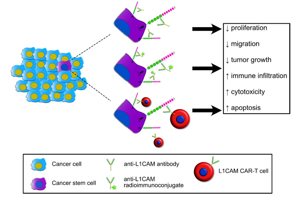 Fig.1 L1CAM is a viable therapeutic target in cancer.1
Service Overview
- WB analysis
- L1CAM single gene analysis
- Flow cytometry and cell sorting
- L1CAM immunoassay
- L1CAM immunohistochemistry.
Workflow
01Sample Processing & Quality Control
We begin with meticulous sample preparation to ensure biomarker integrity. Our team performs nucleic acid and protein isolation, followed by rigorous quality control checks to confirm sample suitability.
02L1CAM Expression Analysis
Using cutting-edge platforms like quantitative PCR (qPCR) for mRNA and ELISA or western blotting for protein, we precisely quantify L1CAM expression levels.
03Functional & Localization Assays
Where required, we perform assays such as immunohistochemistry (IHC) to determine L1CAM's subcellular localization or a cell adhesion assay to assess its functional impact. This step provides critical biological context to the quantitative data.
04Data Analysis & Integration
Raw data from various assays are integrated and analyzed using advanced bioinformatic tools. We perform statistical comparisons, pathway analysis, and generate clear visualizations to highlight significant findings.
05Comprehensive Report Generation
The final step involves compiling all results, analysis, and conclusions into a detailed, easy-to-digest report.
Why Choose Us?
Choosing Creative Biolabs for your L1CAM analysis means partnering with a team that combines decades of scientific expertise with a commitment to excellence. Our specialized focus on cell adhesion molecules, including both L1CAM and ALCAM, provides a unique depth of knowledge that ensures the highest quality results. We understand the nuances of these proteins, from their cleaved soluble forms to their roles in epithelial-to-mesenchymal transition (EMT) and metastasis, enabling a more insightful analysis.
Applications
Oncology Research
Biomarker Discovery & Validation
Identify L1CAM as a potential liquid biopsy marker, especially the soluble form, for early disease detection or monitoring of treatment response.
Therapeutic Development
Support the discovery and validation of novel therapeutic targets by understanding how L1CAM's signaling pathways can be modulated to inhibit cancer progression.
Cell & Molecular Biology Studies
Explore the fundamental biological functions of L1CAM, its interactions with other proteins, and its role in cellular signaling and adhesion in both healthy and diseased states.
FAQs
-
What types of cancer research can benefit most from L1CAM analysis?
Our L1CAM analysis is highly relevant for research into cancers where L1CAM overexpression is common and linked to poor prognosis, such as ovarian, endometrial, colon, and pancreatic cancers. It is particularly valuable for projects focused on biomarker discovery, liquid biopsy development, and targeted therapy research.
-
How does your L1CAM analysis service accommodate different sample types?
We offer a versatile platform that can analyze L1CAM from a wide range of biological materials, including fresh, frozen, and FFPE tissue, as well as serum, plasma, and cell culture lysates. We work with you to determine the most appropriate analytical methods for your specific sample type to ensure optimal results.
-
Can your service help us differentiate between membrane-bound and soluble L1CAM?
Yes, our analysis is designed to distinguish between the full-length, membrane-bound form of L1CAM and its cleaved, soluble ectodomain. This is a critical distinction, as the soluble form is a promising candidate for non-invasive liquid biopsy applications.
-
What are the advantages of using Creative Biolabs for L1CAM analysis instead of performing it in-house?
Our service provides a significant advantage in expertise, equipment, and efficiency. We eliminate the need for you to invest in expensive platforms, develop complex protocols, or handle technical troubleshooting. Our specialists ensure fast, accurate, and reproducible results, allowing your team to focus on core research and development.
By providing high-quality data, expert analysis, and a seamless workflow, Creative Biolabs empowers you to accelerate biomarker discovery, streamline clinical trials, and advance the development of new targeted research. Contact us now!
Reference
- Giordano, Marco, and Ugo Cavallaro. "Different shades of L1CAM in the pathophysiology of cancer stem cells." Journal of clinical medicine 9.5 (2020): 1502. Distributed under Open Access license CC BY 4.0, without modification. https://doi.org/10.3390/jcm9051502
For Research Use Only.