Oxidative Stress Analysis Service
Creative Biolabs provides a wide variety of analysis services for oxidative stress-induced cardiotoxicity to our valued clients. Our team is equipped with extensive expertise and state-of-the-art techniques to accurately evaluate the effects of anticancer agents on heart health, aiding you in identifying and mitigating potential cardiotoxic risks. Moreover, Using a range of highly sensitive testing methods, we can comprehensively assess key indicators such as oxidative stress levels, apoptosis, and myocardial cell damage.
Introduction
Certain anticancer medications can increase the levels of oxygen species (ROS), potentially causing apoptosis and impairing the function of cardiac cells. This oxidative stress not only affects the vital signs of myocardial cells but can also contribute to cardiotoxicity.
Creative Biolabs offers assessments and analyses related to oxidative stress, assisting researchers and companies in evaluating the mechanisms of cardiotoxicity induced by specific anticancer drugs. We utilize a combination of advanced technologies, including high-throughput screening, real-time cell monitoring, and multiple biomarker evaluations, enabling us to accurately capture the effects of oxidative stress on cardiac cells. Through quantitative proteomics and genomics, we delve into the responsive mechanisms of cardiac cells under oxidative stress conditions, unveiling potential toxicity pathways and ensuring the safety of pharmaceuticals.
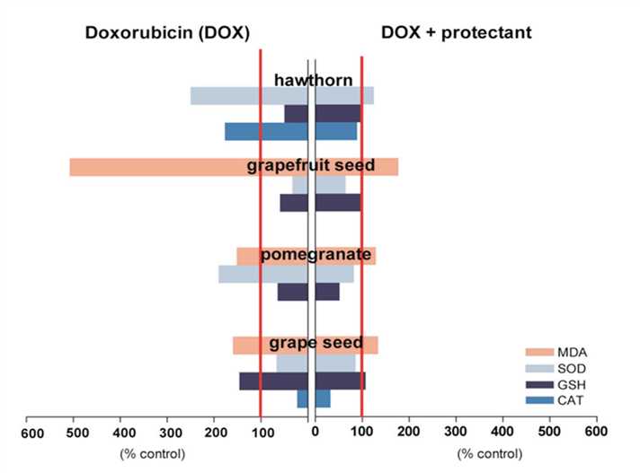 Fig.1 The Oxidative-stress-associated Cardiotoxicity.1,3
Services
Cardiotoxicity resulting from cancer therapies is a significant area of research, with oxidative stress being one of its critical mechanisms.
- Drug Evaluation: Assessing the cardiotoxic effects of anticancer agents through both in vitro and in vivo experiments. Various models, including those using mice or other animal species, can be employed to evaluate the cardiac safety of these drugs.
- Oxidative Stress Biomarker Measurement: Analyzing biomarkers associated with oxidative stress, such as malondialdehyde (MDA), superoxide dismutase (SOD), and glutathione (GSH), to assess the impact of drugs on cardiac cells.
- Cardiac Function Assessment: Utilizing techniques like echocardiography or hemodynamic monitoring to evaluate the effects of anticancer drugs on heart function.
- Molecular Mechanisms Analysis: Examining how anticancer medications trigger oxidative stress through methods like gene expression analysis, proteomics, and metabolomics.
- Free Radical Measurement: Determining levels of free radicals in samples using chemical methods or instrumental techniques, with common approaches including electron spin resonance (ESR) and fluorescence assays.
- Antioxidant Capacity Testing: The antioxidant capacity of biological samples or reagents is evaluated using methods such as the DPPH radical scavenging assay and the ABTS radical scavenging assay.
- Cellular Experiments: These experiments involve cell culture studies to investigate how specific compounds influence oxidative stress levels, with assessments including cell viability, ROS production, and apoptosis rates.
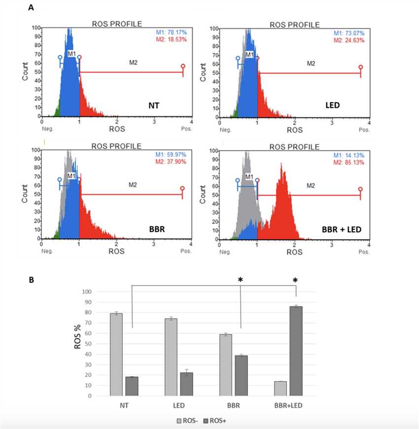 Fig.2 Oxidative Stress Analysis.2,3
Case Study: The mechanism study of cardiotoxicity caused by anticancer drugs through the enhancement of oxidative stress.
1. Selection of Drugs and In Vitro Experiment Design
Choose known or candidate cardiotoxic anticancer agents.
Use cardiac cell lines (like H9c2 or HL-1) or primary cardiomyocytes for the in vitro studies.
2. Assessment of Oxidative Stress Levels
ROS Measurement: Employ fluorescent probes (DCFH-DA) to quantify intracellular reactive oxygen species (ROS).
Antioxidant Enzyme Activity Testing: Measure the activity levels of antioxidant enzymes, including superoxide dismutase, catalase, and glutathione peroxidase.
3. Detection of Apoptosis and Necrosis
Utilize flow cytometry to measure the rate of apoptosis, employing the Annexin V/PI double staining method.
Apply the TUNEL staining technique to assess DNA fragmentation as an indicator of apoptosis.
4. Analysis of Signaling Pathways
Perform Western blotting to analyze the activation status of oxidative stress-related signaling pathways (such as NF-κB, p53, and p38 MAPK).
Conduct qPCR to evaluate changes in the expression levels of relevant genes (like Bax, Bcl-2, and Nrf2).
5. Animal Model Studies
Develop a mouse model and monitor cardiac function and associated oxidative stress markers following drug administration.
Assess changes in cardiac function using echocardiography or small animal MRI.
In the field of cancer drug development, the cardiac toxicity induced by oxidative stress has emerged as a critical issue that demands urgent attention. Creative Biolabs has launched specialized services for analyzing oxidative stress-related cardiotoxicity, providing robust support for drug development and safety assessments. Whether you require data support for basic oxidative stress analysis or need to evaluate the cardiotoxicity of a new agent, we are here to offer you expert services and guidance. Feel free to contact us, and let us help advance your research!
References
- Koss-Mikołajczyk, Izabela, et al. "Natural products counteracting cardiotoxicity during cancer chemotherapy: The special case of doxorubicin, a comprehensive review." International Journal of Molecular Sciences 22.18 (2021): 10037.
- Carriero, Francesca, et al. "Berberine photo-activation potentiates cytotoxicity in human astrocytoma cells through apoptosis induction." Journal of Personalized Medicine 11.10 (2021): 942.
- Distributed under Open Access license CC BY 4.0, without modification.
 Download our brochure