This technique provides insights into the solvent-accessible surface area of a protein, which can be combined with our EM data to pinpoint the exact location of an epitope.
Learn More →B Cell based Electron Microscopy Assay Service
Creative Biolabs' B cell-based electron microscopy assay provides a direct, high-resolution view of B cell and antigen interactions, offering critical insights that go beyond traditional bulk analysis. Our solutions are designed to address key pain points in therapeutic and vaccine development, allowing you to visualize and understand the binding regions of specific antibodies, confirm competing antibodies, and describe mutation maps associated with pathogen escape. This level of detail is essential for a comprehensive understanding of molecular function and disease.
Introduction What We Can Offer Workflow Why Creative Biolabs Customer Reviews FAQs Related Services Contact Us
Electron Microscopy (EM) for Antibody-antigen Complex Analysis
Electron microscopy is a high-resolution method that can characterize the structure, conformational states, and subunit compositions of tissues, cells, and complex macromolecules. Electron microscopy is promising in providing crucial structural information as key proof in analyzing molecular function, cell structure changes, and disease. It can even generate 3D structures on an atomic resolution level using homogeneous, rigid, biological macromolecules. EM can show three-dimensional conformation and molecular interaction style for dynamic, asymmetric biological molecular complexes involved in critical cellular processes.
To learn more about how our capabilities can assist your project, request a consultation.
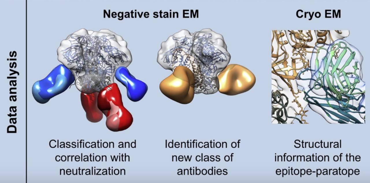 Fig.1 Functions of EM Technologies on epitope analysis. 1
What We Can Offer
Electron Microscope is an advanced technique used for the confirmation of previous results and data on antigen receptor binding domain (RBD) and determinants of antibodies, providing a 3D structure map of the binding reaction of the complex with nanoscale resolution.
| The Function of EM Assay for B Cell Epitope Analysis | Robust EM Platform |
|---|---|
|
|
Electron Microscopy Services at Creative Biolabs
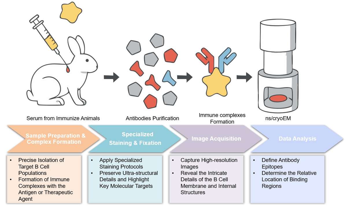
Highlights
Transformative Approach
Our B cell-based electron microscopy assay offers a transformative approach to immunological research. We provide unparalleled resolution and insights that simply aren't possible with conventional methods.
Unparalleled Resolution
This service goes beyond traditional techniques by providing high-resolution images and data at the nano-scale, revealing a level of detail that is essential for understanding complex biological interactions.

Expert Team
Our expertise and cutting-edge technology are the foundation of our service. Our team's deep knowledge and experience ensure that you receive precise and reliable data for your project.
Actionable Insights
We offer a transformative approach to immunological research, providing unparalleled resolution and insights. Our expertise and cutting-edge technology allow us to provide a level of detail that is simply unattainable with conventional methods.
Discover how a partnership with us can streamline your research - Get a quote today.
Customer Reviews
Unprecedented Detail
Using Creative Biolabs' B cell-based electron microscopy assay in our research has significantly improved our understanding of how our lead antibodies bind to their targets. The level of detail we get on epitope mapping is a game-changer for our development pipeline. - Dr. J***s, Biopharma Inc.
Essential for Polyclonal Mapping
The ability to use this assay for mapping polyclonal epitopes was instrumental in our recent HIV immunization study. We were able to follow the development of neutralizing antibodies over time, which confirmed our hypotheses and led to new insights. - Dr. S***h, Academic Research Center.
FAQs
Can your assay be used for my specific therapeutic antibody?
Our assay is highly versatile and can be tailored to various therapeutic antibodies and antigens. By providing us with your specific materials and project goals, we can design a customized workflow to meet your needs and provide the structural insights you require.
How does this assay compare to flow cytometry for B-cell analysis?
While flow cytometry is excellent for sorting large populations of B cells and identifying them by surface markers, it lacks the resolution to visualize the actual binding events and subcellular structures. Our assay provides the detailed, nano-scale visual data that complements flow cytometry, offering a complete picture of cellular interactions.
Related Services
Footprinting Mass Spectrometry
Hydrogen-Deuterium Exchange Mass Spectrometry (HDX-MS)
HDX-MS is used to study the conformational changes and dynamics of proteins, offering a functional complement to the static structural data from EM.
Learn More →X-Ray Crystallography
For ultimate high-resolution atomic structure, X-ray crystallography can be used in conjunction with our EM assay to gain a complete understanding of the epitope-paratope interaction.
Learn More →Antibody Competition Assay
This assay provides functional information on whether multiple antibodies bind to the same or distinct epitopes, which we can then visually confirm with our EM assay.
Learn More →How to Contact Us
Creative Biolabs has developed a reliable T and B-cell assay platform and can also provide assays for monitoring epitope-specific cell responses, but not limited to the above. If you are looking for a cancer epitope discovery service, please look at Creative Biolabs. Do not hesitate anymore and contact us right now.
Reference
- Bianchi, Matteo et al. "Electron-Microscopy-Based Epitope Mapping Defines Specificities of Polyclonal Antibodies Elicited during HIV-1 BG505 Envelope Trimer Immunization." Immunity vol. 49,2 (2018): 288-300.e8. Distributed under an Open Access license CC BY 4.0, without modification. https://doi.org/10.1016/j.immuni.2018.07.009
 Download our brochure
Loading case studies...
Online Inquiry