Electroporation-Mediated DNA Vaccine Delivery for Robust T-Cell & Antibody Responses
Despite their conceptual simplicity, naked plasmid DNA (pDNA) vaccines have historically struggled with poor cellular uptake and limited immunogenicity in large-animal models. Creative Biolabs addresses this bottleneck through an integrated preclinical electroporation (EP) service designed to enhance DNA vaccine delivery by up to 1000-fold compared to direct injection alone. Our platform systematically optimizes the critical EP parameters—field strength, pulse duration, waveform, and electrode geometry—against your specific vaccine construct, injection route, and target tissue. By combining rational plasmid engineering with route-matched EP protocols, we enable researchers to achieve robust, dose-sparing humoral and cellular immune responses that persist through prime-boost regimens. The service spans every stage from construct design through in vivo efficacy validation, giving you a single partner for developing high-performance DNA vaccines against tumor antigens, viral epitopes, and emerging infectious targets.
How EP Transforms Naked pDNA into a Potent Vaccine
The Electrophysical Gate into Target Cells
Electroporation applies brief, controlled electrical pulses at the injection site to transiently destabilize the cell membrane, creating aqueous pores through which plasmid DNA can enter the cytoplasm. Unlike lipid-based transfection or viral vectors, EP is a purely physical method—it requires no additional chemical carriers and works across diverse tissue types including skeletal muscle, dermis, and solid tumor masses. Once inside the cell, the pDNA is trafficked to the nucleus, where the encoded antigen is transcribed, translated, processed via the endogenous antigen-presentation machinery, and displayed on MHC class I molecules. Simultaneously, secreted or cross-presented antigen accesses the MHC class II pathway in professional antigen-presenting cells (APCs). This dual-pathway engagement is the immunological signature that distinguishes EP-delivered DNA vaccines from simple peptide immunization.
EP yields 100- to 1000-fold increases in plasmid delivery and gene expression compared with naked DNA injection. The technique drives antigen expression both in transfected myocytes or keratinocytes at the injection site and in APCs that traffic to draining lymph nodes.
- Core Preclinical Challenges We Address:
- Identifying optimal pulse parameters (voltage, duration, waveform) for each construct and tissue.
- Matching electrode geometry to the injection route (intramuscular, intradermal, intratumoral).
- Quantifying antigen expression kinetics and the resulting antibody and T-cell responses.
- Balancing transfection efficiency with tissue tolerability to preserve animal welfare.
How EP Delivery Compares to Other DNA Administration Routes
| Key Comparison | Naked pDNA Injection Alone | pDNA + Electroporation (EP) |
|---|---|---|
| Gene Expression Level | Low; poor cellular entry of naked plasmid. | 100–1000× higher antigen expression. |
| Antibody Response | Weak or undetectable in large animals. | Robust, sustained humoral immunity with dose sparing. |
| T-Cell Activation | Minimal CD8+ T-cell priming. | Strong CD8+ and CD4+ T-cell responses via cross-presentation. |
| Prime-Boost Flexibility | Limited memory recall. | Excellent prime-boost responses with accelerated recall kinetics. |
End-to-End EP-Mediated DNA Vaccine Development Services
Our preclinical services are structured into flexible, modular packages. Because every plasmid construct and target tissue presents unique electrophysical requirements, all modules can be fully customized—from pulse-parameter matrices to route-specific electrode selection—to align with your vaccine candidate and preclinical model.
Plasmid Construct Engineering
Optimizing the DNA sequence for maximum expression after EP-mediated delivery into the chosen tissue.
- Codon Optimization: Species-specific codon adaptation for the target host.
- Promoter Selection: Strong constitutive promoters (CMV) for high-level antigen expression.
- Antigen Design: Full-length, truncated, or multi-epitope cassettes evaluated in silico.
- Plasmid Backbone: High-yield production vectors with antibiotic-free selection markers.
EP Parameter Matrix Screening
Systematic evaluation of voltage, pulse duration, interval, and waveform to identify the optimal EP protocol for your construct.
- Voltage Titration: Field-strength optimization (V/cm) for each tissue type.
- Waveform Comparison: Square-wave vs. exponential-decay pulse evaluation.
- Pulse-Number Matrix: Testing 2–8 pulses with varied intervals.
- Reporter Quantification: Luciferase or GFP imaging to benchmark expression efficiency.
Electrode Selection & Route Matching
Selection and validation of electrode configurations matched to injection route and target tissue architecture.
- Intramuscular (IM): Needle-array electrodes for deep muscle penetration.
- Intradermal (ID): Surface or minimally invasive electrodes for skin vaccination.
- Intratumoral (IT): Custom geometries for direct tumor electroporation.
- Depot-Type Designs: Electrodes that retain pDNA at the site for extended transfection.
In Vitro & Ex Vivo Expression Verification
Confirming antigen expression before advancing to animal studies using cell-based and tissue-explant assays.
- Cell-Line Transfection: EP-mediated pDNA delivery into cultured myoblasts or epithelial lines.
- Western Blot / ELISA: Quantitative antigen detection in cell lysates and supernatants.
- Flow Cytometry: MHC-I-peptide complex detection on transfected cells.
- Ex Vivo Tissue Imaging: Fluorescent reporter visualization in electroporated tissue sections.
Immunogenicity Assessment
Multi-parameter profiling of the humoral and cellular immune responses induced by EP-delivered DNA vaccines.
- Antibody Titration: Serum IgG, IgG1/IgG2a subclass ELISA for Th1/Th2 bias readout.
- ELISpot / ICS: IFN-γ and granzyme B quantification for antigen-specific T cells.
- Tetramer Staining: Direct enumeration of epitope-specific CD8+ T cells by flow cytometry.
- Dendritic Cell Activation: CD80/86 and MHC-II upregulation in draining lymph node APCs.
In Vivo Efficacy & Tumor Challenge Studies
Definitive proof-of-concept studies in syngeneic or humanized mouse models to demonstrate vaccine-mediated tumor protection.
- Prophylactic Models: Vaccinate → challenge with tumor cells; monitor tumor-free survival.
- Therapeutic Models: Implant tumor → administer EP-DNA vaccine; measure regression.
- Prime-Boost Schedules: Optimize dosing intervals for maximal memory T-cell expansion.
- Combination Studies: EP-DNA vaccine plus immune checkpoint inhibitor co-administration.
Standardized EP-Mediated DNA Vaccine Development Workflow
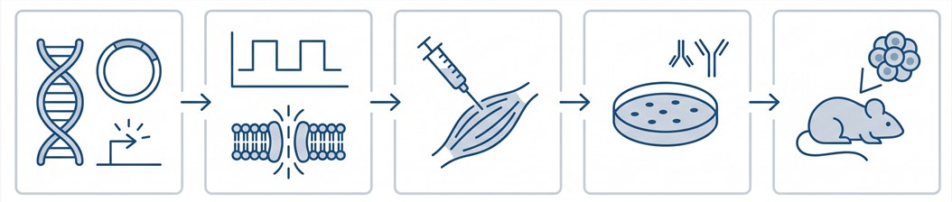
Phase 1 — Plasmid Engineering & Antigen Cassette Design
We design and synthesize the DNA vaccine plasmid, incorporating codon-optimized antigen sequences, high-efficiency promoters, and optimized Kozak sequences. Each construct undergoes sequence verification and endotoxin-free preparation at research-grade scale.
Core Platforms Enabling High-Efficiency EP Delivery
Why Choose Creative Biolabs?
We don't apply a one-size-fits-all protocol. Each construct-tissue combination is evaluated through a purpose-built matrix to identify optimal transfection conditions.
Deep experience with intramuscular, intradermal, and intratumoral EP, including route-specific electrode selection and pulse-parameter tuning.
Antibody titer, subclass bias, ELISpot, tetramer staining, and cytokine profiling are bundled into each study—no need to outsource immunomonitoring.
From codon optimization through tumor-challenge endpoints, every step is handled within a single integrated workflow for full traceability.
Research Insight: EP as a Transformative DNA Vaccine Delivery Tool
Key Findings from Published Preclinical Studies
Electroporation-mediated DNA vaccine delivery has been validated across multiple preclinical models, demonstrating consistent advantages in antigen expression, immune response magnitude, and tumor protection relative to naked plasmid injection.
-
100–1000× Expression Boost: A comprehensive review of next-generation DNA vaccine delivery systems confirmed that EP achieves 100- to 1000-fold increases in plasmid-driven antigen expression compared with direct intramuscular or intradermal injection. This expression enhancement is the mechanistic foundation for the superior immunogenicity observed across viral, bacterial, and tumor-antigen vaccine candidates.
-
Route-Dependent Immune Polarization: Studies in non-human primates demonstrated that intradermal EP not only enhanced antigen expression in skin-resident APCs but also acted as a natural adjuvant, promoting dendritic cell maturation and lymph node homing without exogenous immunostimulatory molecules. The route of EP delivery—intramuscular, intradermal, or intratumoral—strongly influences the balance between humoral and cellular immunity.
-
Parameter Optimization Drives Translation: Systematic optimization of pulse parameters and electrode geometries has been shown to restore vaccine efficacy even in immunosenescent models, and recent advances in device miniaturization are expanding the repertoire of accessible target tissues for preclinical investigation.
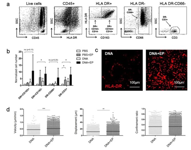
Fig.1 Characterization of skin immune infiltrates following EP or DNA + EP treatment.2,5