p62 (SQSTM1) DNA Vaccine Platform for Broad-Spectrum Cancer Immunotherapy
Creative Biolabs provides a specialized preclinical platform for the development of DNA vaccines encoding p62 (SQSTM1), an autophagy-associated protein that meets the stringent criteria of an ideal tumor antigen: strong immunogenicity, tumor-essential function yet dispensable for normal tissues, and overexpression in at least 10 distinct human cancer types. Our p62-encoding DNA vaccine development service leverages a unique dual-form antigen strategy — co-delivering protease-resistant and protease-susceptible p62 variants within a single plasmid — to maximize both MHC class I and class II antigen presentation, driving coordinated CD8+ and CD4+ T cell responses alongside robust humoral immunity. Validated across syngeneic mouse and rat models of melanoma, lung cancer, breast carcinoma, and sarcoma, our p62 vaccine platform has demonstrated consistent tumor growth inhibition (3- to 4-fold) and strong suppression of spontaneous and induced metastasis. By integrating plasmid engineering, codon optimization, and preclinical efficacy testing, we empower researchers to advance next-generation immunotherapy candidates targeting a tumor antigen that is both broadly relevant and mechanistically compelling.
Why p62 (SQSTM1) Stands Out as a Cancer Vaccine Antigen
A Unique Autophagy-Informed Tumor Antigen
p62 (sequestosome-1), encoded by the SQSTM1 gene, is a multidomain scaffold protein central to selective autophagy and oncogenic signaling. Unlike most tumor-associated antigens (TAAs) that are lineage-restricted or mutation-dependent, p62 accumulates aberrantly across a wide spectrum of malignancies due to its dual role as both an autophagy substrate and a signaling amplifier. In tumor cells, oncogenic drivers such as RAS, PIK3CA, and HER2 converge on the p62-KEAP1-NRF2 axis, creating a sustained positive-feedback loop that drives p62 overexpression — a phenomenon documented in breast, lung, colon, liver, pancreatic, prostate, glioblastoma, melanoma, renal, and myeloma tumors. This widespread dysregulation positions p62 as a uniquely pan-cancer antigen with broad preclinical applicability.
DNA immunization ensures intracellular antigen expression, routing p62 through the endogenous MHC-I processing pathway for CD8+ T cell priming while simultaneously releasing antigen for cross-presentation. Encoding two antigen forms — one protease-resistant for sustained MHC-I loading and one susceptible for cross-presentation — amplifies both arms of the adaptive response beyond what single-form or peptide vaccines can achieve.
- Core Preclinical Challenges We Address:
- Confirming p62 overexpression status in client tumor models before vaccine design.
- Engineering dual-form p62 antigen cassettes with balanced protease susceptibility.
- Quantifying coordinated CD8+ and CD4+ T cell responses in vitro and in vivo.
- Evaluating anti-metastatic efficacy across spontaneous and induced metastasis models.
p62 DNA Vaccine vs. Conventional Single-Antigen Plasmid Vaccines
| Key Comparison | Conventional Single-Antigen DNA Vaccines | p62 DNA Vaccine Platform |
|---|---|---|
| Antigen Scope | Lineage-specific; often restricted to 1–2 tumor types. | Pan-cancer antigen overexpressed in 10+ human tumor types. |
| Antigen Processing Strategy | Single-form antigen; biased MHC pathway utilization. | Dual-form antigen: protease-resistant + susceptible variants for balanced MHC-I/II. |
| Safety Profile | Varies by antigen; potential for on-target off-tumor toxicity. | p62 dispensable for normal tissues; no on-target organ toxicity observed. |
| Anti-Metastatic Activity | Often limited to primary tumor control. | Validated suppression of spontaneous and induced metastasis across 3 models. |
Preclinical p62 DNA Vaccine Development Service Modules
Our service portfolio is organized into modular, independently configurable packages. We understand that every tumor indication presents unique p62 expression profiles and immunological contexts; therefore, all modules can be customized — from codon-optimized plasmid architecture to model-specific efficacy endpoints — to align with your preclinical development goals.
p62 Expression & Antigen Validation
Comprehensive profiling of p62/SQSTM1 in your tumor model to confirm candidacy and guide construct design.
- Expression Profiling: Immunoblotting and immunohistochemistry for p62 levels in tumor vs. normal tissue.
- Mutation Analysis: Screening of SQSTM1 coding sequence for tumor-specific variants or truncations.
- Autophagy Status: Assessment of autophagic flux (LC3-I/II ratio) to contextualize p62 accumulation mechanism.
- GATA4 Pathway Readout: Evaluation of p62-GATA4 senescence axis in your tumor model.
Dual-Form Antigen Cassette Engineering
Rational design of the p62 open reading frame to encode both protease-resistant and protease-susceptible antigen forms.
- Codon Optimization: Species-specific codon adaptation for maximum expression in the target host.
- Degron Engineering: Strategic placement of ubiquitination signals to tune proteasomal degradation rate.
- Kozak & UTR Optimization: 5' and 3' UTR design for enhanced translation initiation and mRNA stability.
- Subcellular Routing: Optional signal peptide engineering for secretory or membrane-targeted p62 expression.
Plasmid Construction & QC
Endotoxin-free plasmid DNA production with rigorous quality control for preclinical immunization studies.
- Vector Backbone Selection: CMV or hybrid promoter-driven expression cassettes with optimized polyadenylation signals.
- Endotoxin-Free Production: Large-scale plasmid purification with < 0.1 EU/mg endotoxin specification.
- Sequence Verification: Full-plasmid Sanger sequencing confirming ORF integrity and regulatory element placement.
- Transfection Validation: Confirmation of p62 protein expression by western blot in transfected mammalian cells.
In Vitro Antigen Processing & Presentation
Functional validation of dual-form p62 antigen processing through both MHC class I and class II pathways.
- MHC-I Presentation Assay: Co-culture of p62-transfected cells with antigen-specific CD8+ T cell hybridomas.
- Cross-Presentation Readout: Dendritic cell uptake and MHC-II presentation of secreted p62 forms.
- Proteasome Dependence: Confirmation of dual-form processing using proteasome and lysosomal inhibitors.
- Dendritic Cell Maturation: Assessment of co-stimulatory marker upregulation (CD80, CD86) upon p62 antigen encounter.
Preclinical Immunogenicity Profiling
Multi-parameter assessment of vaccine-induced T cell, B cell, and cytokine responses.
- Anti-p62 Antibody Titer: ELISA-based quantification of p62-specific IgG responses over time.
- T Cell ELISpot: IFN-γ and IL-4 ELISpot assays measuring antigen-specific CD8+ and CD4+ T cell frequencies.
- Intracellular Cytokine Staining: Multiparametric flow cytometry for IFN-γ, TNF-α, and IL-2 co-expression.
- T Cell Proliferation: CFSE dilution assays quantifying p62-specific T cell expansion in vitro.
In Vivo Efficacy & Anti-Metastatic Evaluation
Comprehensive preclinical efficacy package including primary tumor growth, metastasis, and survival endpoints.
- Tumor Growth Inhibition: Serial caliper measurement of subcutaneous tumors in syngeneic mouse or rat models.
- Metastasis Quantification: Enumeration of spontaneous lung/liver metastases and induced metastasis (intravenous challenge) models.
- Survival Analysis: Kaplan-Meier survival curves with median survival comparison between vaccine and control groups.
- Tumor Microenvironment Profiling: IHC and flow cytometry for TIL density, Treg/FoxP3, and myeloid cell composition.
p62 DNA Vaccine Development Workflow
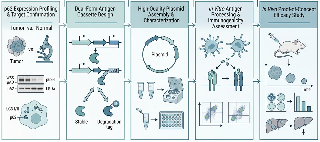
Phase 1 — p62 Expression Profiling & Target Confirmation
We perform quantitative analysis of p62 protein levels in your tumor tissue versus matched normal tissue by immunoblotting and IHC. Autophagic flux status (LC3-I/II, p62 aggregate formation) is assessed to confirm that p62 accumulation is driven by oncogenic signaling rather than autophagy deficiency alone, establishing a mechanistic rationale for vaccine targeting.
Enabling Technology Platforms for p62 DNA Vaccine Development
Why Choose Creative Biolabs for p62 DNA Vaccine Development?
Our scientists bring specialized knowledge of the p62/SQSTM1 signaling network, including the p62-KEAP1-NRF2 and p62-GATA4 axes, enabling rational antigen design beyond generic plasmid construction.
Our p62 vaccine platform is benchmarked against published preclinical datasets demonstrating consistent tumor growth inhibition (3 to 4-fold) and metastasis suppression across melanoma, lung, breast, and sarcoma models.
Unlike single-form plasmid vaccines, our co-expression approach ensures simultaneous MHC-I and MHC-II engagement, generating the coordinated CD8+ and CD4+ T cell responses critical for durable anti-tumor immunity.
Every service module is independently configurable with clearly defined milestones, timelines, and deliverables. You receive raw data alongside processed reports — no black-box results.
Research Insight: p62 DNA Vaccine Efficacy Across Multiple Preclinical Models
Key Findings from Preclinical p62 Vaccine Studies
The p62-encoding DNA vaccine has demonstrated broad-spectrum preclinical activity, with consistent anti-tumor and anti-metastatic effects observed across multiple independent studies encompassing five transplantable tumor models in mice and rats.
-
Pan-Cancer Tumor Growth Suppression: p62 DNA vaccination achieved 3-fold tumor growth inhibition in B16 melanoma and Lewis lung carcinoma, and 4-fold inhibition in S37 sarcoma, demonstrating activity independent of tumor histology or tissue of origin.
-
Metastasis Suppression Across Routes: The vaccine reduced spontaneous lung metastasis (LLC model) by 4-fold, regional lymph node metastasis by 6-fold (S37 model), and intravenous-induced metastasis (B16 model) by 4-fold, suggesting both colonization and outgrowth are affected.
-
Safety Validated Across Species: Comprehensive toxicology evaluations in rats, mice, guinea pigs, and dogs revealed no acute or chronic toxicity, no hematological or organ-level pathology, and no impairment of B or T cell function — supporting a favorable preclinical safety profile.
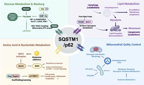
Fig.1 p62-mediated context-dependent oncogenic adaptation across tumor types.2,4