
EpCAM (KSA) Peptide & DC Cancer Vaccine Development
Creative Biolabs provides comprehensive preclinical development services for KSA-based cancer vaccines, covering antigen characterization, epitope mapping, anti-idiotypic antibody design, peptide and protein vaccine formulation, dendritic cell (DC)-based loading, in vitro immunogenicity assessment, and in vivo efficacy evaluation. KSA, also known as epithelial glycoprotein (EGP) or epithelial cell adhesion molecule (EpCAM), is a 40 kDa transmembrane glycoprotein overexpressed on the surface of most adenocarcinomas—including colorectal, gastric, pancreatic, breast, lung, and prostate cancers. This solution is designed for researchers developing tumor-associated antigen (TAA) vaccines, validating KSA-specific T-cell responses, optimizing anti-idiotypic strategies, or evaluating combination approaches with immune checkpoint inhibitors. Our scientists can provide customized study designs, assay development, quality control, and integrated preclinical packages tailored to your tumor indication, antigen format, and translational goals.
KSA (EpCAM): A Compelling TAA Target for Adenocarcinoma Vaccines
From Adhesion Molecule to Vaccine Antigen
KSA (EpCAM) is a type I transmembrane glycoprotein belonging to a family of epithelial cell surface antigens homologous to nidogen, a matrix adhesion protein of neuroectodermal origin. In normal epithelial tissues, KSA is expressed in a polarized manner at the basolateral membrane, where it contributes to cell-cell adhesion and maintains tissue architecture. Upon malignant transformation, this polarized distribution is lost; KSA becomes homogeneously distributed across the entire tumor cell membrane, mirroring the expression pattern changes observed with carcinoembryonic antigen (CEA). This redistribution exposes previously sequestered epitopes to immune surveillance, making KSA an attractive target for active immunotherapy.
KSA is overexpressed in over 90% of colorectal and gastric adenocarcinomas, and its expression is retained in metastatic lesions and circulating tumor cells. Anti-KSA antibodies have been detected in patients following anti-idiotypic antibody vaccination, confirming that KSA is immunogenic in humans without detectable toxicity to normal tissues.
- Core Preclinical Challenges We Address:
- Selecting immunodominant KSA epitopes restricted by common HLA alleles.
- Breaking tolerance to a self-antigen through optimized adjuvant and carrier strategies.
- Designing anti-idiotypic antibodies that mimic KSA conformational epitopes.
- Distinguishing anti-tumor immunity from on-target, off-tumor reactivity in vivo.
KSA Expression: Normal Epithelium vs. Adenocarcinoma
| Key Comparison | Normal Epithelial Tissue | Adenocarcinoma (KSA-Positive) |
|---|---|---|
| Membrane Distribution | Polarized; restricted to basolateral surface. | Homogeneous; circumferential membrane staining. |
| Expression Level | Moderate; varies by tissue type. | Markedly upregulated; retained in metastases. |
| Epitope Accessibility | Sequestered by tight junctions; limited immune access. | Fully exposed to antibody and T-cell recognition. |
| Cancer Stem Cell Association | Not applicable. | Enriched on CSC populations; targets tumor-initiating cells. |
End-to-End KSA Vaccine Service Packages
Our preclinical services are structured into flexible, modular packages. We understand that every project is unique; therefore, all modules can be fully customized—from epitope selection to vaccine formulation and animal model choice—to align with your therapeutic goals and tumor indications.
Antigen Evaluation & Epitope Mapping
Comprehensive analysis of KSA antigenic structure to identify the most immunogenic epitopes for vaccine design.
- Expression Profiling: Verification of KSA expression levels across target adenocarcinoma cell lines and patient-derived samples.
- Epitope Prediction: Bioinformatics-driven identification of HLA-A2 and other allele-restricted KSA peptide epitopes.
- Binding Affinity: Peptide-T2 binding assays to rank epitopes by MHC class I binding strength.
- Conformational Mapping: Identification of discontinuous B-cell epitopes recognized by anti-KSA monoclonal antibodies (17-1A, KS1/4).
Anti-Idiotypic Antibody Design
Development of anti-idiotypic antibodies that function as internal images of KSA, enabling active immunization without administering the tumor antigen itself.
- Mimotope Selection: Screening phage display libraries for antibody fragments that mimic KSA conformational epitopes.
- Cross-Reactivity: Validation that anti-idiotypic antibodies elicit sera capable of binding native KSA on tumor cells.
- Immunogenicity Screening: In vitro B-cell activation assays to confirm humoral response induction.
- Carrier Optimization: Conjugation to carrier proteins (KLH, CRM197) to enhance T-helper dependent responses.
Vaccine Construct Production
Synthesis and assembly of KSA vaccine candidates across multiple platforms tailored to your immunization strategy.
- Peptide Vaccines: Production of multi-epitope synthetic long peptide (SLP) pools spanning immunodominant KSA regions.
- Protein Vaccines: Recombinant KSA ectodomain production in mammalian or insect expression systems.
- DNA/RNA Vaccines: Construction of plasmid or mRNA vectors encoding KSA for endogenous antigen expression.
- Viral Vectors: Generation of recombinant viral vectors (adenoviral, vaccinia-based) carrying KSA transgenes.
DC-Based KSA Vaccine Formulation
Specialized dendritic cell loading and maturation protocols to maximize KSA-specific T-cell priming.
- Peptide Pulsing: Optimization of KSA peptide loading onto monocyte-derived DCs for MHC class I and II presentation.
- mRNA Electroporation: Transfection of DCs with KSA-encoding mRNA for sustained endogenous antigen processing.
- Maturation Cocktails: TLR agonist and cytokine combinations to drive CD80/CD86 co-stimulatory marker expression.
- Adjuvant Screening: Evaluation of immunostimulatory adjuvants to break self-tolerance and enhance immunogenicity.
Comprehensive Potency Evaluation
Multi-level assessment of vaccine-induced humoral and cellular anti-tumor immune responses.
- Humoral Assays: ELISA and flow cytometry to quantify anti-KSA antibody titers and tumor cell surface binding.
- Cellular Assays: ELISpot (IFN-γ), intracellular cytokine staining, and DC-T co-culture cytotoxicity assays.
- In Vivo POC: Efficacy studies in syngeneic or humanized mouse models bearing KSA-expressing tumors.
- Immune Profiling: Tumor-infiltrating lymphocyte analysis, cytokine monitoring, and survival tracking.
QC & Translational Data Packages
Quality assurance and comprehensive documentation to support translational advancement of KSA vaccine candidates.
- Antigen Characterization: Purity, endotoxin, and stability testing of vaccine constructs.
- DC Phenotyping: Flow cytometry verification of CD11c, HLA-DR, CD80, CD83, and CD86 expression.
- Lot Consistency: Reproducibility assessment across independent vaccine preparations.
- Safety Screening: Preliminary toxicology and off-target reactivity assessments in relevant in vitro panels.
Preclinical KSA Vaccine Development Workflow
Phase 1 — KSA Antigen Characterization & Epitope Selection
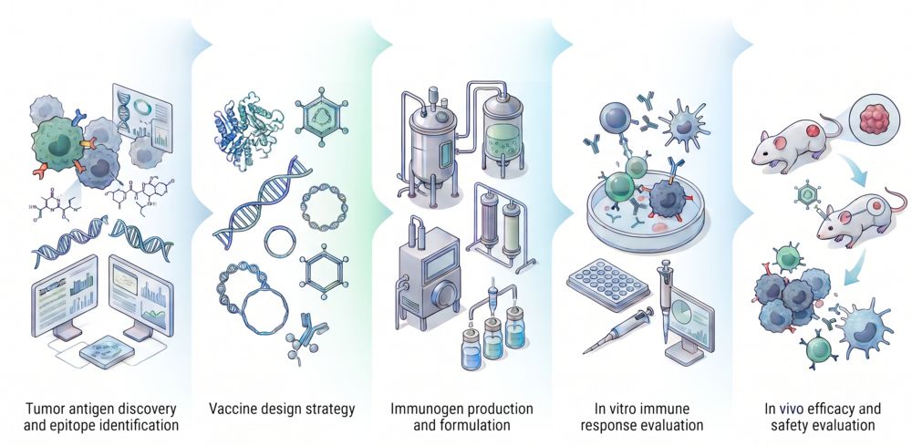
We begin by confirming KSA (EpCAM) expression on your target adenocarcinoma cell lines or patient-derived xenograft samples. Using bioinformatics prediction tools and peptide-T2 binding assays, we identify HLA-A2-restricted KSA peptide epitopes with high MHC binding affinity. Conformational B-cell epitopes recognized by established anti-KSA monoclonal antibodies (17-1A, KS1/4) are also mapped to guide anti-idiotypic antibody design.
Enabling Technologies for KSA Vaccine Development
Why Choose Creative Biolabs?
Years of focused experience in tumor-associated antigen vaccine development, with particular strength in epithelial antigens including KSA, CEA, and MUC1.
From peptide and protein vaccines to DC-based formulations and anti-idiotypic approaches, we offer the full spectrum of vaccine modalities under one roof.
Our assay development team builds project-specific readouts—ELISpot, cytotoxicity, antibody binding—that precisely measure KSA-directed immune responses.
From antigen evaluation through in vivo efficacy, we provide a single integrated workflow with rigorous QC, complete documentation, and dedicated scientific communication.
Research Insight: EpCAM Peptide-DC Vaccination in Hepatocellular Carcinoma
Key Findings from Preclinical Studies
EpCAM (KSA) peptide-primed dendritic cell vaccination has demonstrated significant anti-tumor immunity in preclinical hepatocellular carcinoma models, providing a strong rationale for expanding KSA-targeted vaccine strategies across adenocarcinoma indications.
-
Efficient CTL Induction: EpCAM peptides with high HLA-A2 binding affinity, when loaded onto monocyte-derived DCs, generated potent cytotoxic T lymphocytes that specifically killed EpCAM+ HepG2 tumor cells in vitro.1
-
In Vivo Tumor Growth Inhibition: Vaccination of tumor-bearing mice with EpCAM peptide-CTLs significantly delayed HepG2 tumor growth, with reduced EpCAM expression in residual tumor tissue confirmed by immunohistochemistry.1
-
Bispecific T-Cell Engager Synergy: Oncolytic vaccinia virus engineered to promote EpCAM-directed T-cell engagement enhanced intratumoral CD8+ T-cell infiltration and reduced T-cell exhaustion, supporting its potential synergy with KSA vaccine strategies.2
-
Broad Adenocarcinoma Applicability: EpCAM-targeted T-cell–engaging immunotherapies have been investigated across multiple epithelial cancer types, including pancreatic, colorectal, and gastric adenocarcinomas, supporting KSA as a broadly relevant target across adenocarcinomas.
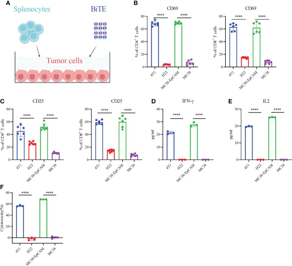
Fig.1 Secreted EpCAM-targeted T-cell–engaging molecules activate naïve T cells and induce cytotoxicity against tumor cells.2, 3