Visualize Terminal Glycans Using Fluorescence or SDS-PAGE
In recent years, glycoprotein detection has attracted great attention. As an expert in this field, Creative Biolabs has completed many challenging projects and has won a good reputation among global customers.
The analysis of glycans is essential to better understand their functions in biological processes. A variety of glycan detection technologies have been developed. Among the various technologies, fluorescence detection and sodium dodecyl sulfate-polyacrylamide gel electrophoresis (SDS-PAGE) are the most commonly used methods for visualizing terminal glycans.
Introduction of SDS-PAGE
SDS-PAGE is often used to determine the molecular weight of proteins in biomedical research. Mainly in the presence of sodium lauryl sulfate by polyacrylamide gel electrophoresis to distinguish proteins of different molecular weights. It can also be used to visualize terminal glycans. It is worth noting that this technique requires the protein studied on the gel and the protein standard used for calibration to adopt the same shapes after SDS treatment.
Application and Examples of Fluorescence Detection
Fluorescence is a common detection technique. Due to its simplicity and high sensitivity, fluorescence plays an irreplaceable role in optical microscopy, flow cytometry, chromatography, immunodiagnosis, electrophoresis, and DNA sequencing.
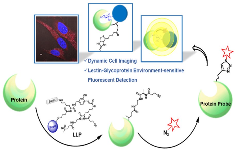 Fig.1 Analysis of lectin-glycoprotein interactions by fluorescent labeling.1, 2
This technology can perform spatial observation well and judge the location of the target protein, but it is not suitable for large flow detection. Fluorescence microscopes have different magnification objectives. High magnification objectives are used to observe fine structures while small magnification objectives are suitable for a small number of statistics.
Flow cytometry is a precision instrument that can analyze complex target groups in detail in a short time. With the development of technology, fluorescent microspheres combined with biomarkers and analyzed on flow cytometer instruments provide a new method for the detection platform. It is worth noting that this technology plays an important role in the quantitative detection of galactomannan.
Basic Features of Fluorescence Detection
The technology has a self-checking function to avoid the output of wrong data.
All detection processes are closed-tube operations. Fluorescence is detected at the reaction tube to effectively prevent contamination and solve the most difficult problem in PCR technology.
The method can meet the clinical needs of quantification.
Take advantage of the sensitivity of spectral signals to effectively improve detection sensitivity.
Non-toxic testing throughout the process.
Save the tedious process of post-processing.
Print the results directly without recording a large amount of data.
The two technologies mentioned above focus on different directions in glycoprotein determination. In the actual application process, different methods can be combined for visualizing terminal glycans. We have decades of experience in glycoprotein analysis and detection. Our scientists can choose the appropriate detection method to meet your different needs. Please contact us for more details.
References
-
Li, Yiran, et al. "Cell-surface glycan labeling and sensing." Targets 2.1 (2023): 1-31.
-
Under Open Access license CC BY 4.0, without modification.
For Research Use Only.
Resources
