Glycosylation Analysis Service for Ebola Virus Glycoprotein
Ebola Virus Glycoprotein
Ebola virus (EBOV) is a single-stranded, membrane-enveloped RNA virus and belongs to the Filoviridae family. It triggers deadly viral hemorrhagic fevers in humans. EBOV has a genome of 18 kb, encoding seven genes: NP, VP35, VP40, GP, VP30, VP24, and L2. The entry of EBOV requires its envelope glycoprotein GP, which is made up of GP1 and GP2 subunits. GP1 is involved in receptor binding and comprises four domains: the base, receptor-binding domain (RBD), glycan cap, and mucin-like domain (MLD). GP2 is essential for membrane fusion and is highly conserved among filoviruses. EBOV GP is heavily glycosylated, including N-linked glycans and variable O-linked glycans in the MLD. C-Mannosylation is also found at one site within the GP. These glycans play important roles in host cell attachment, viral stability, fusion with host cell membranes, and immune evasion. For example, high-mannoseglycans mediate the EBOV attachment to host cells through lectins DC-SIGN/L-SIGN and protect GP from neutralizing antibodies. Understanding the glycosylation patterns of EBOV proteins, particularly GP, is essential for developing vaccines and therapeutic treatments.
Glycosylation Analysis Services for EBOV Glycoprotein at Creative Biolabs
Armed with years of experience and cutting-edge technologies in Glycoprotein Analysis, Creative Biolabs offers a comprehensive panel of services for characterizing glycosylation patterns. We have developed viral glycoproteomics services designed for sensitive detection and in-depth analysis of viral glycosylation sites and attached glycan patterns, including N-linked, O-linked, and C-linked glycosylation profiles for GP from various EBOV strains and subtypes. EBOV GP has been reported to contain 11–17 N-glycosylation sites, and over 80 predicted O-glycosylation sites, more than 50 different N-glycan structures were identified. Our services extend to offering comprehensive characterizations of glycosylation site distributions within EBOV GP. It is noteworthy that these distributions can significantly differ among subtypes. By evaluating the distribution of different glycans in different EBOV strains, we aim to unravel the complexities of glycosylation patterns and their role in EBOV viral biology and contribute to the development of vaccine and antiviral treatment.
Our Platform for Glycosylation Analysis of EBOV GP
Our approaches to achieving in-depth glycoproteomic characterization and high sequence coverage involve the digestion of different proteases during sample preparation, combined with advanced mass spectrometry platforms like MALDI-TOF/TOF MS (CID/HCD/ETD). We have also developed different glycan and glycopeptide analysis software and used multiple strategies to increase confidence in structure assignments. Furthermore, we perform comparative analyses of glycosylation patterns among different pathogenic viruses and host cells. These optimized strategies enable comprehensive mapping of glycosylation sites in EBOV GP and glycan profiling at each site, including both composition and structure analysis.
Glycosylation of EBOV GP as the Target of Antiviral Therapeutics
High-mannose glycans present on EBOV GP play an important role in viral entry and infection, targeting these specific glycans is essential for the development of potential antiviral treatments. Mannose-binding lectin (MBL) therapy, using high doses of MBL, binds to EBOV and leads to virus neutralization through complement activation, highlighting the significance of viral glycosylation in therapy.
Advantages of Our Services
-
Cutting-edge technologies and the latest equipment for MS
-
In-depth characterization and high-confidence data analysis
-
Comprehensive profiling of glycosylation sites and glycan structures
-
Comparative glycosylation analysis among different virus strains
-
Experienced professionals specializing in glycosylation analysis
Published Data
Technology: MS-based site-specific glycosylation analysis
Journal: Communications Biology
IF: 6.548
Published: 2022
Results: The researchers employed glycoproteomics to characterize the site-specific glycosylation of EBOV GP. This analysis revealed the presence of 16 unique O-linked glycosylation sites in the MLD and identified complex glycosylation patterns at most of the N-linked sites, with notable enrichment of unprocessed glycans at conserved sites. Additionally, the study confirmed the presence of C-mannosylation at the W288 residue in full-length trimeric GP.
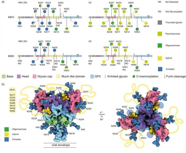 Fig.1 N-linked glycosylation profiling of EBOV GP.1, 2
Creative Biolabs provides specialized detection and analysis services to decipher the glycosylation patterns of EBOV glycoprotein, offering valuable insights into viral infection mechanisms and vaccine development. If you have specific needs related to viral glycoprotein analysis, please contact us for more detailed information.
References
-
Peng, Weiwei, et al. "Glycan shield of the ebolavirus envelope glycoprotein GP." Communications Biology 5.1 (2022): 785.
-
Under Open Access license CC BY 4.0, without modification.
For Research Use Only.
Related Services
