
Gene Therapy for Respiratory Genetic Disorders
Gene therapy can address respiratory genetic disorders at their molecular source by restoring, replacing, editing, or regulating disease-associated genes in relevant airway or lung cells. Its potential extends to conditions such as cystic fibrosis, alpha-1 antitrypsin deficiency, and primary ciliary dyskinesia, but durable benefit depends on overcoming mucus barriers, achieving efficient cell-specific delivery, managing immune responses, and maintaining expression in renewing respiratory tissues.
Introduction
Gene therapy for respiratory genetic disorders offers promising approaches to address disease-causing mutations through gene addition, RNA replacement, genome editing, and other targeted strategies. This resource discusses airway delivery, disease-relevant models, efficacy assessment, and key translational considerations. Creative Biolabs supports related research through gene therapy development for genetic disorders, including vector design, delivery strategy development, model construction, and analytical evaluation.
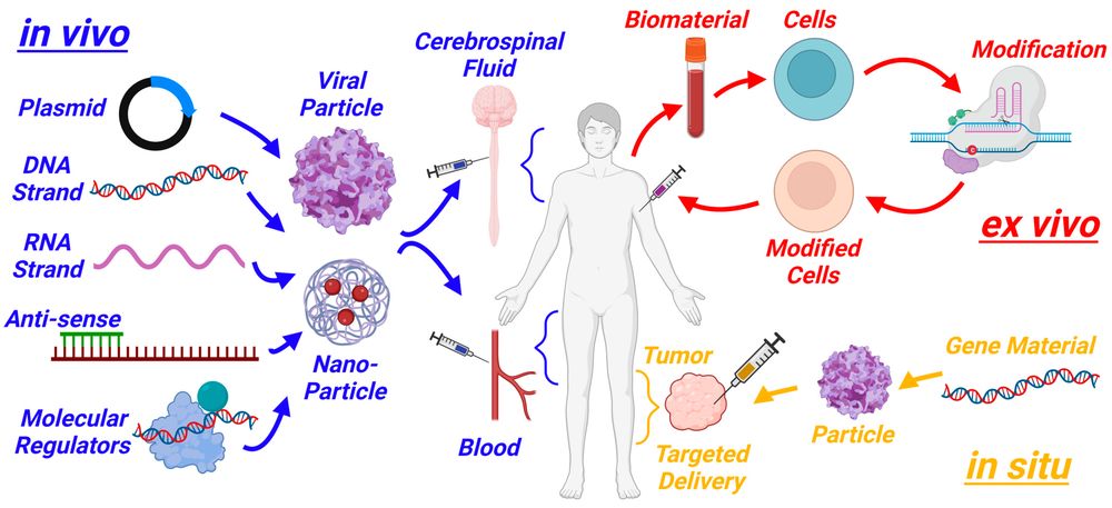 Figure 1. Schematic of three gene therapy approaches: in vivo, ex vivo, and in situ.1
Why the Respiratory Tract Is a Distinct Target?
The epithelial surface is accessible but strongly defended
Inhaled delivery can place a payload near diseased airway cells without first crossing every systemic barrier. Yet the same surface is designed to exclude foreign material. Mucus, mucociliary clearance, cough, surfactant, extracellular nucleases, antimicrobial factors, resident phagocytes, and tight epithelial junctions can reduce residence or entry. Chronic infection and inflammation change mucus rheology, receptor expression, epithelial integrity, and immune tone. A formulation or vector that works in a healthy model may therefore behave differently in diseased human airways.
Cell identity and epithelial renewal determine durability
Secretory, ciliated, basal, club, alveolar type I and II, and other pulmonary cells contribute different functions. Correcting a mature cell can produce rapid activity but may fade as the epithelium turns over. Correcting a stem or progenitor cell could support durable descendants, but those cells may be difficult to access from the lumen and may require controlled integration or editing. The necessary fraction of corrected cells is disease specific and depends on whether function is cell autonomous, shared across the surface, or supplied as a secreted factor.
Disease-Specific Resource Pathways
Gene Therapy for Alpha-1 Antitrypsin Deficiency
Gene Therapy for Alpha-1 Antitrypsin Deficiency should be planned around two linked but non-identical disease mechanisms. Loss of circulating functional alpha-1 antitrypsin reduces control of neutrophil elastase in the lung, while selected mutant proteins, especially Z-alpha-1 antitrypsin, can polymerize and accumulate in hepatocytes. Gene addition may raise a functional secreted protein without removing mutant protein; transcript silencing may reduce liver toxicity while worsening systemic deficiency unless function is replaced; editing may address both but requires efficient and well-characterized hepatocyte correction. Readouts should include functional antiprotease activity, serum exposure, pulmonary protection, intracellular polymers, liver injury, and durability.
Gene Therapy for Primary Ciliary Dyskinesia
Gene Therapy for Primary Ciliary Dyskinesia focuses on restoring the molecular machinery of motile cilia in airway epithelial cells. Primary ciliary dyskinesia (PCD) is genetically heterogeneous, and variants can affect dynein arms, radial spokes, central-pair components, microtubule organization, or assembly factors. Payload size and mutation class vary widely. Protein expression alone is insufficient: a useful intervention must restore correct axonemal localization, beat frequency or waveform, coordinated mucociliary transport, and epithelial health. Transient messenger RNA replacement, viral or non-viral gene addition, genome editing, and corrected airway-cell approaches answer different durability and delivery questions.
Gene Therapy for Cystic Fibrosis
| Disorder | Primary Defect | Potential Genetic Objective | Defining Functional Readout |
|---|---|---|---|
| Alpha-1 antitrypsin deficiency | Low functional circulating AAT; mutant protein may accumulate in liver | Add functional SERPINA1, silence mutant RNA, or correct the locus | Active AAT and protease control plus hepatic polymer burden |
| Primary ciliary dyskinesia | Defective motile-cilia structure, assembly, or movement | Replace transcript or gene, correct variant, or restore airway cells | Ciliary waveform and mucociliary transport |
| Cystic fibrosis | Insufficient CFTR ion-channel function | Add CFTR, correct selected variants, or modulate RNA | CFTR-dependent ion transport and airway-surface function |
Choose Strategy by Biological Defect
The same platform should not be forced across disorders. Strategy selection begins with whether the disease is a loss of function, toxic gain of function, dominant-negative process, splicing defect, or combination. Payload size, required expression level, target-cell lifespan, mutation diversity, and acceptable redosing then narrow the options. AAV-mediated gene addition can provide durable episomal expression in selected cells, while genome editing may preserve endogenous control but adds editing efficiency and by-product questions.
- Gene addition is attractive when a functional coding sequence can fit the vector and expression does not need to reproduce every endogenous regulatory feature.
- Messenger RNA replacement is non-integrating and adaptable but usually transient, making aerosol tolerability and repeat dosing central.
- Genome editing can repair, disable, or rewrite an allele, yet delivery of editor components and characterization of on-target products are major burdens.
- Transcript silencing can reduce a toxic product but may require simultaneous replacement when the normal protein is also required.
- Ex vivo corrected airway cells offer extensive pre-administration testing but face engraftment, distribution, epithelial integration, and scale barriers.
- Combination strategies should be justified by separate evidence for each component and by assays that identify which active species produces benefit.
The Airway Delivery Chain
A respiratory product succeeds only if every link from aerosol generation to intracellular action remains adequate. Measuring deposited dose alone cannot distinguish mucus trapping from target-cell entry, and measuring tissue DNA or RNA cannot prove functional correction.
- Generate an aerosol or local dose with a stable particle or vector distribution that preserves biological activity during nebulization and handling.
- Reach the intended airway region with a device, breathing pattern, particle size, and dose volume suited to the model and eventual population.
- Cross or avoid mucus and extracellular barriers without producing damaging surfactant disruption, aggregation, or excessive innate immune activation.
- Bind and enter the relevant epithelial population; use cell-resolved evidence rather than whole-lung concentration alone.
- Escape endosomal or other intracellular sequestration when required, then reach the nucleus, cytosol, or apical membrane according to payload mechanism.
- Produce the correct protein, RNA, or edit at sufficient magnitude and duration to restore a disease-specific function.
- Maintain a tolerable pulmonary and systemic profile across the intended dose interval, including repeat administration when durability is limited.
Models That Preserve Respiratory Biology
Rodents can support mechanism, distribution, dose range, and inflammatory screening, but airway geometry and cell tropism differ from humans. Ferret, pig, sheep, or non-human primate models may better represent selected aspects of airway size, mucus, delivery devices, or immune response, yet each introduces ethical, cost, species-reagent, and translatability constraints. Gene-edited disease models can provide causal genotypes, but the model should reproduce the functional endpoint the therapy intends to restore.
| Research Question | Model Fit | Priority Readouts | Important Limitation |
|---|---|---|---|
| Does payload reach the target epithelial cell? | Human air-liquid interface culture with apical dosing | Cell type, intracellular location, intact payload, expression | Lacks whole-lung deposition and systemic exposure |
| Does correction restore disease function? | Patient-derived and isogenic airway models | CFTR transport, ciliary motion, mucus transport, AAT function as relevant | Donor and differentiation variability |
| Can the aerosol reach the intended region? | Large-animal device and deposition study | Regional recovery, imaging, cell-resolved biodistribution | Species anatomy and breathing still differ from humans |
| Is repeat dosing tolerable? | Disease-relevant airway culture plus selected animal model | Barrier, cytokines, histology, antibodies, activity over cycles | Chronic infection context may be incompletely modeled |
A Measurement Hierarchy for Respiratory Rescue
Evidence should be layered so a negative functional result can be localized to delivery, expression, molecular activity, or biology. A positive distal endpoint is also more credible when supported by proximal steps.
- Delivery: emitted dose, aerodynamic distribution, regional deposition, residence, intact payload, and target-cell uptake.
- Molecular action: vector genome or editor exposure, RNA, protein abundance, localization, editing spectrum, or splice product.
- Cell function: CFTR-dependent ion transport, ciliary beat and transport, active AAT secretion, or another disease-defining assay.
- Tissue function: airway-surface hydration, mucus transport, epithelial integrity, inflammatory tone, infection-relevant response, or protease balance.
- Durability: persistence during epithelial turnover, repeat-dose response, recovery after dosing stops, and resistance or immunity that changes later doses.
- Safety: local histology, barrier injury, cytokines, immune responses, ectopic distribution, systemic exposure, and platform-specific liabilities.
Near-Term Research Priorities
Progress is likely to come from better alignment between disease biology and delivery rather than from one universal lung vector. Modular payloads and standardized functional assays can make genotype-specific programs more efficient, while human airway models can expose species and disease-state failures earlier. The most useful innovation is one that closes a measured gap in the delivery-to-function chain.
- Develop vectors and non-viral carriers with defined tropism for disease-relevant airway populations and measurable repeat-dose performance.
- Use aerosol devices and formulations that preserve payload activity and can be bridged from model to human airway geometry.
- Establish shared thresholds linking corrected cell fraction or molecular activity to CFTR, ciliary, mucus, or antiprotease function.
- Improve editing methods that minimize persistent nuclease exposure and characterize complete on-target outcomes in airway progenitors and mature cells.
Frequently Asked Questions
Q: Why is inhaled gene therapy difficult even though the airway surface is directly accessible?
A: The airway is protected by mucus, clearance, cough, extracellular enzymes, immune surveillance, and epithelial barriers. Disease-related infection and inflammation can further change deposition, uptake, and tolerability.
Q: Do all respiratory genetic disorders require gene delivery directly to the lung?
A: No. Alpha-1 antitrypsin deficiency can be approached through liver-directed production or correction because the protective protein is mainly produced in hepatocytes and then circulates to the lung.
Q: What is the difference between gene addition and genome editing in the airway?
A: Gene addition supplies a functional expression cassette without necessarily changing the endogenous locus. Genome editing changes a genomic sequence but requires efficient delivery and characterization of on-target and off-target products.
Q: Which airway cells should be targeted?
A: The answer depends on disease and durability. Mature ciliated or secretory cells may provide rapid function, while basal or other progenitor cells may support longer-lived corrected descendants but are harder to access.
Q: How should efficacy be measured for primary ciliary dyskinesia?
A: Measure correct ciliary protein localization, beat frequency and waveform, coordinated mucociliary transport, epithelial health, and durability. Expression alone does not establish functional rescue.
Q: Can CFTR expression alone prove that a cystic fibrosis gene therapy works?
A: No. Expression should be connected to apical localization, CFTR-dependent chloride or bicarbonate transport, airway-surface and mucus function, and a tolerable pulmonary profile.
Published Data
Case 1: CRISPR Adenine Base Editing Corrects Nonsense Mutations in Cystic Fibrosis Organoid Biobank
This study demonstrates the functional repair of cystic fibrosis (CF) nonsense mutations using CRISPR adenine base editors (ABEs) in patient-derived intestinal organoids. Cystic fibrosis is caused by mutations in the CFTR gene, with premature termination codons (PTCs) preventing full-length functional protein expression. Researchers applied ABEs to precisely convert targeted A·T base pairs to G·C in a CF organoid biobank harboring diverse CFTR nonsense mutations. Without generating double-stranded DNA breaks or relying on donor templates, ABE treatment efficiently corrected the stop codons, restoring full-length CFTR protein expression. Functional swelling assays in response to forskolin confirmed the full restoration of CFTR anion channel activity in edited organoids. This study provides crucial proof-of-concept for precision base editing in respiratory and systemic genetic diseases, demonstrating that ABEs can permanently correct disease-causing point mutations and recover organ-level tissue function in primary human stem cell models.
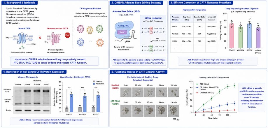 Figure 2. Adenine base editing repairs CF nonsense mutations in patient-derived organoids.
Overview of What Creative Biolabs Can Provide
Creative Biolabs can support research-stage respiratory genetic therapy programs with vector and payload design, airway-targeting strategies, disease-relevant model construction, and analytical evaluation. The services below were selected from the supplied GT Services inventory because they map directly to the delivery, correction, model, and potency decisions described in this resource.
| Research Need | Related Creative Biolabs Support | How It Connects to the Current Resource Topic |
|---|---|---|
| Design a disease-matched viral vector | Custom Viral Vector Development | Supports vector choice and construction around payload size, target cell, duration, route, and safety question. |
| Develop an AAV platform for gene therapy | Adeno-associated Virus Vector Development Service | Provides an AAV development route when episomal expression and airway or liver tropism are appropriate. |
| Improve tissue or cell targeting | Tissue/Cell Specific Targeting Advanced Adeno-Associated Virus Vector Service | Addresses capsid and targeting choices needed to reach specific respiratory or hepatic cell populations. |
| Design an AAV gene-addition cassette | AAV Design for Gene Addition | Supports functional gene expression for loss-of-function programs such as CFTR or selected ciliary genes. |
| Develop an AAV editing vector | AAV Vector Design for Gene Editing | Supports delivery design for mutation-specific correction while recognizing payload and exposure constraints. |
| Build gene-edited disease models | Disease Modeling related Gene Editing Service | Creates causal or corrected cell systems for airway mechanism and rescue studies. |
| Analyze vector quality and function | Viral Vector Analysis | Connects identity, titer, purity, safety, and potency attributes to respiratory study performance. |
| Establish mechanism-linked vector potency | Potency of Viral Vector | Supports assays based on expression, transport, ciliary function, or another disease-relevant endpoint. |
To discuss a respiratory target, airway delivery barrier, disease model, or functional evidence plan, contact us today to connect with our scientific team.
References
- Henderson M L, Zieba J K, Li X, et al. Gene therapy for genetic syndromes: understanding the current state to guide future care. BioTech, 2024, 13(1): 1. https://doi.org/10.3390/biotech13010001 Distributed under Open Access license CC BY 4.0, with modification.
- Geurts M H, de Poel E, Amatngalim G D, et al. CRISPR-based adenine editors correct nonsense mutations in a cystic fibrosis organoid biobank. Cell stem cell, 2020, 26(4): 503-510. e7. 10.1016/j.stem.2020.01.019
