
Gene Therapy for Hematologic Genetic Disorders
Gene therapy can provide durable treatment for inherited hematologic disorders by correcting disease-causing mutations, restoring functional gene expression, or modifying a patient's hematopoietic stem cells. Approaches based on viral vector delivery or genome editing are being developed for hemophilia, sickle cell disease, Wiskott–Aldrich syndrome, and severe combined immunodeficiency. Successful development requires careful control of targeting efficiency, engraftment, expression durability, off-target effects, immunogenicity, and manufacturing consistency.
Introduction
Gene therapy for hematologic genetic disorders uses in vivo gene transfer or ex vivo modification of hematopoietic stem and progenitor cells to restore missing blood proteins or correct inherited defects in blood and immune lineages. Creative Biolabs discusses strategy selection, therapeutic evaluation, durability, and safety in this page. Creative Biolabs also supports related programs through the development of gene therapy for hematopoietic disorders, including vector design, HSPC-focused development, and analytical planning.
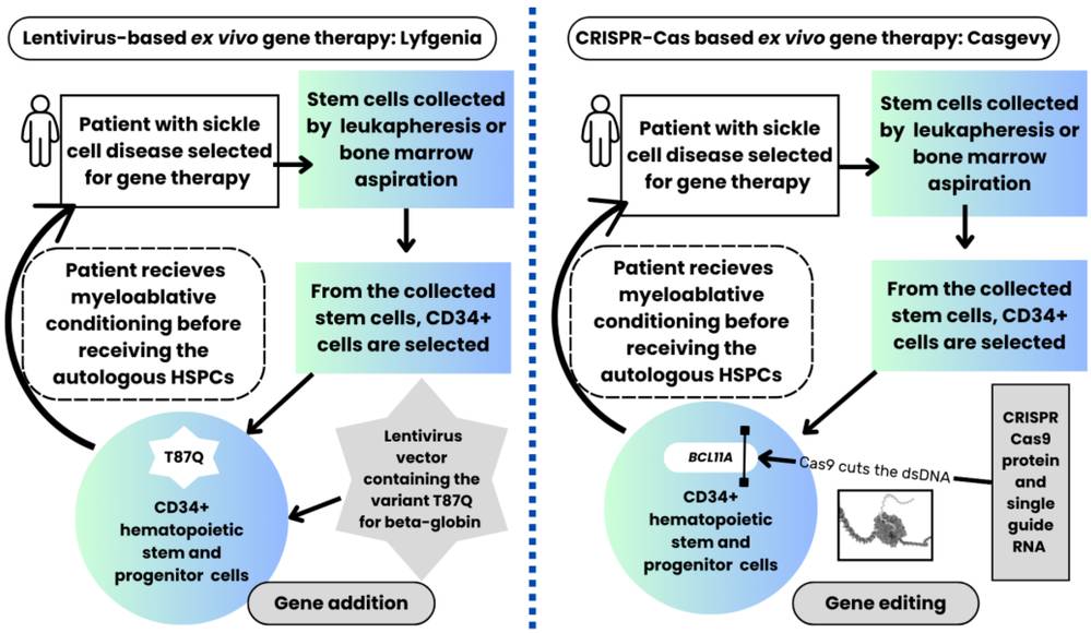 Figure 1. Gene therapy process in patients with sickle cell disease receiving the FDA-approved lentiviral vector-based gene therapy (left side of the image) compared with the CRISPR-Cas9-based gene therapy (right side).1
Disease-Specific Resource Pathways
Gene Therapy for Hemophilia
Gene therapy for hemophilia aims to establish sustained factor VIII or IX activity and reduce bleeding and replacement-factor use. In vivo AAV gene addition has produced approved products for selected patients, while eligibility, neutralizing antibodies, liver health, vector dose, steroid use, durability, and long-term follow-up remain important. See the related hemophilia A/B research pathway for focused vector-development context.
Gene Therapy for Sickle Cell Disease
Gene therapy for sickle cell disease can add an anti-sickling globin or edit regulatory DNA to increase fetal hemoglobin. Both strategies typically modify autologous CD34-positive HSPCs ex vivo and require myeloablative conditioning before reinfusion. A rise in fetal hemoglobin or vector marking should be connected to durable multilineage engraftment and reduction of disease-relevant events. Lentiviral HSPC development for sickle cell disease is one established gene-addition route alongside genome editing.
Gene Therapy for Wiskott-Aldrich Syndrome
Wiskott-Aldrich syndrome (WAS) is caused by pathogenic variants in WAS and affects platelets and multiple immune lineages. Autologous HSPC gene addition seeks regulated WAS protein expression across myeloid and lymphoid descendants while avoiding the genotoxicity seen with some earlier vector designs. Because too little or inappropriate expression may leave lineage-specific defects, potency should include functional cytoskeletal or immune assays rather than protein presence alone. Lentiviral development for Wiskott-Aldrich syndrome requires lineage-aware evidence.
Gene Therapy for SCID
SCID is a family of disorders rather than one molecular target. X-linked SCID involves IL2RG, ADA-SCID involves adenosine deaminase deficiency, and other forms affect signaling, recombination, metabolism, or lymphocyte development. The required corrected lineage, selective advantage, conditioning intensity, and functional readout differ by genotype. X-SCID lentiviral vector development should not be generalized automatically to every SCID genotype.
In Vivo Protein Production vs. Ex Vivo HSPC Engineering
The two dominant development architectures distribute risk differently. In vivo delivery packages the therapeutic cassette and vector as the administered product; dose, tropism, immunity, organ safety, and expression durability are inseparable from the patient. Ex vivo HSPC therapy creates a living product outside the body; collection, cell quality, vector or editing reagents, culture, cryopreservation, release testing, conditioning, and engraftment all contribute to outcome. Ex vivo control allows extensive characterization before infusion but cannot fully predict long-term repopulation. In vivo treatment avoids cell collection and manufacturing of an autologous cell product but provides less opportunity to remove or rework exposed cells.
| Development Feature | In Vivo Gene Delivery | Ex Vivo HSPC Engineering | Interpretive Consequence |
|---|---|---|---|
| Primary product | Vector or non-viral delivery system carrying a genetic payload | Genetically modified autologous HSPC population | Release tests and dose units are fundamentally different. |
| Targeting problem | Reach a production organ or target cell after administration | Modify repopulating cells, then achieve homing and engraftment | Pre-infusion modification does not prove in vivo persistence. |
| Durability source | Vector persistence and stable expression in target tissue | Self-renewal and multilineage output of corrected HSPCs | Time-to-failure mechanisms differ by platform. |
| Major exposure risk | Off-target organs, immunity, dose-related toxicity, and shedding | Conditioning, culture-related fitness, integration or edit risk, and clonal behavior | Safety panels must be platform-specific. |
| Retreatment | May be limited by antibodies, organ exposure, or vector class | May require recollection, remanufacture, and repeated conditioning | Repeat therapy cannot be assumed for either route. |
The Ex Vivo HSPC Chain
Define the corrected cell population, relevant lineages, minimum long-term marking or editing, and functional threshold before mobilization and collection.
Qualify starting material for identity, viability, stem/progenitor composition, disease-related fitness, prior treatment effects, and manufacturing suitability.
Optimize vector exposure or editing to balance on-target modification with viability, stemness, genomic integrity, vector-copy distribution, and process duration.
Demonstrate potency with proximal molecular evidence and a function linked to the affected lineage, not only a bulk percentage or surface marker.
Control cryopreservation, thaw, transport, dose calculation, and infusion readiness because post-thaw recovery influences the effective cell dose.
Match conditioning to the engraftment requirement and patient context, then track marrow recovery, lineage reconstitution, function, clonality, and late events.
Safety Questions Differ by Platform
Safety cannot be reduced to a generic gene-therapy checklist. AAV hemophilia programs emphasize infusion reactions, neutralizing antibodies, liver enzymes, hepatocyte exposure, transgene-product immunity, dose-related findings, shedding, and long-term expression. Integrating lentiviral HSPC products require replication-competent lentivirus testing, vector-copy and insertion-site characterization, clonal surveillance, and assessment of promoter or transgene effects. Genome-edited HSPCs add off-target changes, large deletions, translocations, chromosomal abnormalities, p53-related selection, and unintended functional consequences. Ex vivo programs also carry collection, mobilization, conditioning, infection, infertility, organ toxicity, and engraftment risks that are not caused solely by the genetic modification.
| Risk Domain | AAV In Vivo Program | LV HSPC Program | Edited HSPC Program |
|---|---|---|---|
| Immune response | Capsid and transgene antibodies, cellular immunity, infusion effects | Immune recovery, transgene immunity, infection during conditioning | Editor or delivery immunity plus immune recovery and infection |
| Genomic risk | Persistence and rare integration-related questions | Integration-site distribution and clonal expansion | Off-target edits, structural variants, translocations, and clonal selection |
| Organ/process toxicity | Liver and systemic dose-related findings | Mobilization, culture, cryopreservation, conditioning, and engraftment | Editing process, culture, conditioning, and engraftment |
| Long-term monitoring | Expression durability, liver status, malignancy signals, and shedding as relevant | Lineage marking, clonality, malignancy, and function | Edit persistence, clonality, malignancy, and function across lineages |
Current Landscape of Gene Therapy for Hematologic Genetic Disorders
As of August 2026, approved genetic therapies exist for selected hemophilia and sickle cell populations, and authorized therapies also exist for Wiskott-Aldrich syndrome and ADA-SCID in relevant jurisdictions. These milestones do not make the diseases or platforms interchangeable. Eligibility, genotype, age, organ function, donor options, neutralizing antibodies, conditioning tolerance, manufacturing access, long-term safety, and regional labeling all shape use. Product approvals should be verified against current regulatory records before clinical claims are published. A research resource should explain mechanism and development evidence without offering treatment advice or implying that an experimental construct is equivalent to an authorized product.
- Do not extrapolate an approved vector, editor, promoter, or conditioning regimen to a different disease without new biological and product evidence.
- Separate clinical benefit demonstrated by a product from the general promise of its platform class.
- Account for regional regulatory status, indication wording, age, genotype, eligibility testing, and required long-term follow-up.
- Treat conditioning burden, fertility, collection feasibility, and treatment-center capability as part of the therapeutic strategy.
- Preserve disease-specific functional endpoints even when manufacturing and analytical platforms are shared.
How to Choose a Development Path?
A development path should start with the functional deficit and the cell population capable of correcting it. A secreted factor deficiency may favor in vivo production if a safe organ can provide sufficient regulated output. A cell-intrinsic defect favors ex vivo HSPC engineering when corrected cells can engraft and repopulate the required lineages. Gene addition is efficient when regulated cDNA expression is acceptable; editing is attractive when endogenous control, a regulatory switch, or allele-specific correction is important. The decision should include a fallback if efficiency, cell fitness, payload size, immunity, conditioning, or durability misses the target.
- Choose in vivo gene addition when a production organ can supply a circulating factor and target-organ exposure has a plausible safety margin.
- Choose ex vivo HSPC gene addition when stable multilineage expression is required and an integrating cassette can be regulated appropriately.
- Choose editing when the disease mechanism benefits from endogenous regulation, precise correction, or a validated regulatory disruption and genomic risks can be characterized.
- Use transplant or disease models that test long-term repopulating cells, not only short-lived progenitors or transformed cell lines.
- Define stop criteria for inadequate function, loss of stem-cell fitness, excessive vector copy, genotoxic findings, poor engraftment, or an impractical conditioning requirement.
Frequently Asked Questions
Q: Why can hemophilia use liver-directed gene therapy when bleeding occurs throughout the body?
A: The liver can produce and secrete coagulation factor into circulation. The therapeutic target is therefore a competent production cell, while the functional effect is systemic hemostasis.
Q: Why are hematopoietic stem cells used for sickle cell disease, Wiskott-Aldrich syndrome, and SCID?
A: HSPCs self-renew and generate multiple blood and immune lineages. Correcting long-term repopulating cells can provide durable descendants carrying the therapeutic change.
Q: What is the difference between HSPC gene addition and genome editing?
A: Gene addition introduces a functional cassette, often with an integrating lentiviral vector. Editing changes endogenous or regulatory DNA. They differ in expression control, efficiency, genomic risks, analytics, and potency strategy.
Q: Does a high editing or transduction percentage guarantee durable benefit?
A: No. Bulk measurements may overrepresent short-lived cells. Durability depends on modification of long-term repopulating HSPCs, cell fitness, engraftment, multilineage output, and disease-relevant function.
Q: Why is conditioning used before autologous HSPC gene therapy?
A: Conditioning creates marrow space and supports engraftment of modified cells. Its intensity and toxicity are major parts of the risk-benefit profile and may differ by disease and product.
Q: Are gene therapies approved for all four disease groups discussed here?
A: Approved or authorized products exist for selected hemophilia, sickle cell disease, Wiskott-Aldrich syndrome, and ADA-SCID settings, but status, indication, eligibility, and availability differ by jurisdiction and change over time.
Q: Which long-term safety signals are monitored after HSPC gene therapy?
A: Monitoring may include blood counts, lineage marking or editing, insertion sites, clonal diversity, malignancy, autoimmunity, infections, organ effects, reproductive risks, durability, and need for supportive treatment.
Published Data
Case 1: CRISPR-Cas9 Gene Editing for Sickle Cell Disease and Transfusion-Dependent β-Thalassemia
This study provides early clinical proof-of-concept for ex vivo CRISPR-Cas9 gene editing in autologous CD34+ hematopoietic stem and progenitor cells (HSPCs) to treat transfusion-dependent β-thalassemia (TDT) and sickle cell disease (SCD).
Researchers used electroporation to deliver CRISPR-Cas9 targeting the erythroid-specific enhancer of the BCL11A gene (~80% allele editing without off-target effects). Disrupting this enhancer downregulates BCL11A—a repressor of γ-globin—thereby reactivating fetal hemoglobin (HbF) expression. Following myeloablative conditioning and autologous cell reinfusion, both patients maintained high levels of edited alleles and pancellular HbF elevation for over a year. Consequently, the TDT patient achieved complete transfusion independence, while the SCD patient experienced total elimination of vaso-occlusive crises. This study demonstrates that CRISPR-Cas9 disruption of the BCL11A enhancer effectively reactivates HbF, offering a transformative cure for major hemoglobinopathies.
Overview of What Creative Biolabs Can Provide
Creative Biolabs can support research-stage hematologic gene therapy programs with disease-matched viral vector design, HSPC-focused lentiviral development, and platform-specific analytical planning. The Services pages below were selected from the supplied GT inventory to connect distinct protein-deficiency and blood-cell correction questions to relevant research capabilities.
| Research Need | Related Creative Biolabs Support | How It Connects to the Current Resource Topic |
|---|---|---|
| Design a disease-matched viral vector | Custom Viral Vector Development | Supports payload, vector, regulatory cassette, route, duration, and comparator choices for hematologic research. |
| Develop an AAV platform for gene therapy | Adeno-associated Virus Vector Development Service | Provides an in vivo vector path for secreted-protein strategies such as selected coagulation-factor programs. |
| Develop an AAV strategy for hemophilia | Hemophilia A/B | Addresses factor VIII or IX cassette, expression, activity, and disease-focused vector questions. |
| Design lentiviral vectors for HSPC gene therapy | Lentiviral Vector Design for Hematopoietic Stem Cell-based Gene Therapy Development | Supports stable gene transfer while considering HSPC transduction, expression control, and cell fitness. |
| Develop an HSC-based lentiviral gene therapy | Lentiviral Vector Development for Hematopoietic Stem Cell-based Gene Therapy | Connects vector design to autologous HSC modification, engraftment, and multilineage durability. |
| Develop lentiviral vectors for sickle cell disease | Lentiviral Vector Development for Sickle Cell Disease | Supports anti-sickling globin and HSPC gene-addition research with disease-relevant functional evidence. |
| Develop lentiviral vectors for Wiskott-Aldrich syndrome | Lentiviral Vector Development for Wiskott-Aldrich Syndrome | Focuses regulated WAS expression across immune and platelet lineages and associated safety questions. |
| Develop lentiviral vectors for SCID | Lentiviral Vector Development for X-SCID | Provides a subtype-specific route for IL2RG-focused HSPC gene-addition research rather than a generic SCID claim. |
To discuss a hematologic defect, in vivo or HSPC strategy, disease model, or evidence plan, contact us today to connect with our scientific team.
References
- Kansal R. Curing sickle cell disease by allogeneic hematopoietic stem cell (HSC) transplantation toward in vivo HSC gene therapy. Genes, 2025, 16(11): 1367. https://doi.org/10.3390/genes16111367 Distributed under Open Access license CC BY 4.0, with modification.
- Frangoul H, Altshuler D, Cappellini M D, et al. CRISPR-Cas9 gene editing for sickle cell disease and β-thalassemia. New England Journal of Medicine, 2021, 384(3): 252-260. 10.1056/NEJMoa2031054.
