Tissue-Targeted Lentiviral Vectors
Introduction
Tissue-targeted lentiviral vectors (LV) are designed to improve where a lentiviral payload enters, where it is expressed, or both. In practice, targeting is rarely controlled by a single feature. Envelope pseudotyping can influence entry, promoter choice can restrict transcription, miRNA target sites can suppress expression in off-target cells, and the route or model system can determine exposure. This resource explains how these layers work together and why tissue targeting should be validated as a multi-readout research question.
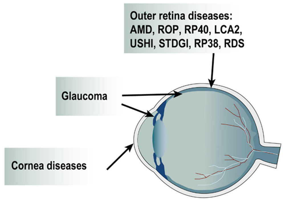 Figure 1. Diseases currently targeted by LV.1,3
Targeting Is a Layered Design Problem
A tissue-targeted lentiviral vector should be defined by the biological decision it is meant to control. Some projects need selective entry; others only need selective expression after broad entry has occurred.
| Targeting Layer | Primary Question | Typical Design Tools |
|---|---|---|
| Particle entry | Which cells receive the vector particle? | Envelope pseudotyping, ligand retargeting, route and dose control |
| Transcription | Which transduced cells express the payload? | Tissue-specific promoters, enhancers, promoter size optimization |
| Post-transcriptional detargeting | Which cells suppress the transcript? | miRNA target sites in untranslated regions |
| Functional restriction | Where does the delivered biology produce a useful effect? | Inducible elements, payload localization, assay timing |
Entry Targeting through Pseudotyping
Pseudotyping can alter the receptor interactions and membrane-fusion behavior of lentiviral particles. This strategy is powerful, but it must be tested in the correct target-cell state because receptor expression can change with differentiation, activation, inflammation, or culture conditions.
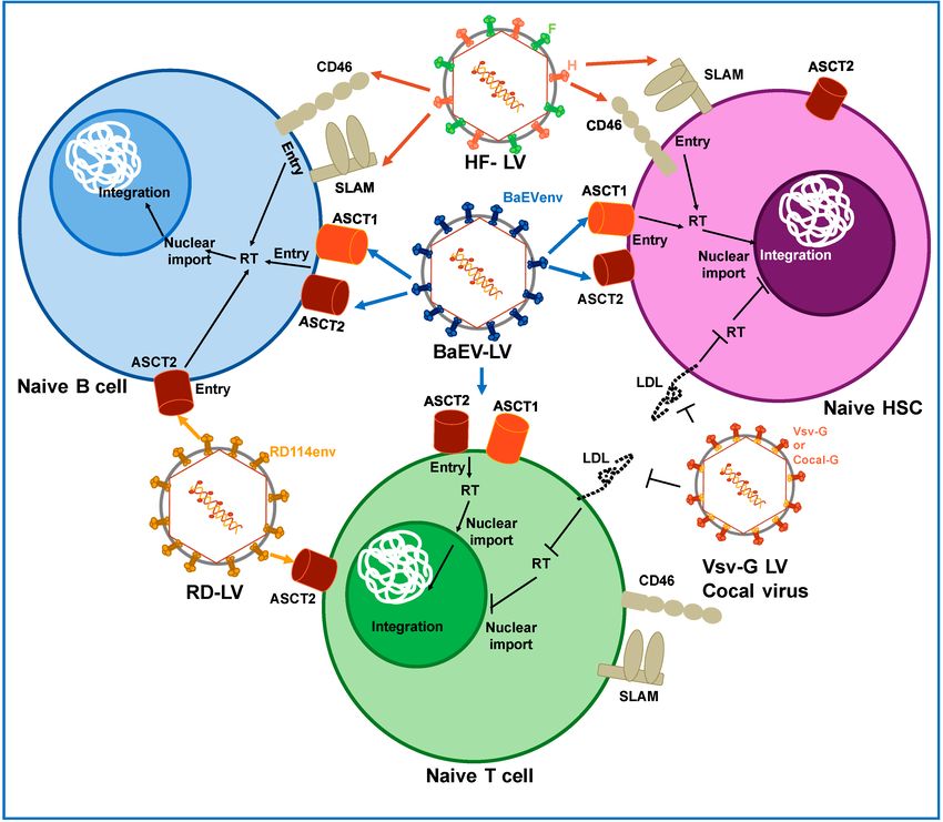 Figure 2. Pseudotyping of lentiviral vectors. Lentiviral vectors were generated expressing different envelopes from other viruses such as measles virus (HF), baboon envelope (BaEV) and feline endogenous retrovirus RD114 envelope gps (RD).2,3
Envelope choice and receptor context
- Hepatocyte-oriented pseudotyping should be evaluated against liver-relevant cells and off-target cell types, not only one permissive line.
- Neuronal-entry strategies require attention to axonal access, differentiation stage, and receptor distribution.
- Lung-cell and myocyte entry should be interpreted in models that capture epithelial or muscle-specific receptor context.
Ligand and antibody-based retargeting
- Ligand-guided entry can increase selectivity when a surface marker is strongly enriched on the target cell.
- Binding is not enough; fusion competence, particle incorporation, receptor internalization, and steric accessibility also need testing.
- Retargeted particles should be compared with a broad-entry control and an envelope-negative or irrelevant-target control.
Expression Targeting after Transduction
When broad or partially broad entry is acceptable, expression targeting can provide another level of specificity. This is especially useful when the target tissue is difficult to reach with a perfectly selective envelope.
- Tissue-specific promoter control can restrict transcription to cells that contain the required transcription-factor environment.
- miRNA-based detargeting can reduce expression in off-target lineages that express a selected microRNA.
- Inducible expression modules can add timing control when constitutive expression would confound the biology.
| Strategy | Best Use | Main Risk |
|---|---|---|
| Tissue-specific promoter | Restrict expression after entry into mixed-cell populations | Lower expression strength compared with viral promoters |
| miRNA target sites | Suppress transcripts in defined off-target cells | Incomplete suppression if miRNA expression is variable |
| Ligand retargeting | Improve particle entry into marker-positive cells | Can reduce titer or fusion efficiency |
| Inducible system | Control timing or dose of transgene expression | Requires inducer-response validation and leakiness testing |
Validation Should Separate Entry from Expression
A common mistake is to measure only the final transgene signal and call the vector targeted. That signal can reflect entry, transcription, transcript stability, translation, selection, or cell survival.
Recommended readout logic
- Measure vector genomes or early reporter signal to estimate entry or delivery.
- Measure mRNA and protein output to evaluate expression control.
- Compare target cells with credible off-target cells under matched multiplicity and culture conditions.
- Use physical and functional titer data so that apparent specificity is not simply caused by lower vector input.
Model selection
- Two-dimensional cell lines are useful for first-pass comparisons but often overestimate targeting performance.
- Primary cells, co-cultures, organoids, or tissue slices can reveal off-target entry and promoter leakiness.
- In vivo work should match route, dose, immune context, and biodistribution endpoints to the targeting claim.
Design Trade-offs in Tissue-Targeted Lentiviral Vectors
More targeting layers can increase specificity, but they also increase engineering complexity. The best design is the simplest design that answers the research question with adequate specificity and measurable controls.
| Trade-off | Why It Matters | Practical Mitigation |
|---|---|---|
| Specificity vs. titer | Highly selective envelopes can reduce particle yield or infectivity | Screen several envelope formats and normalize by functional titer |
| Promoter strength vs. restriction | Native tissue promoters may be weaker than viral promoters | Compare enhancer variants and minimum-expression thresholds |
| Detargeting vs. payload stability | miRNA sites can reduce expression where the miRNA is present | Map miRNA expression in target and off-target cells |
| Model relevance vs. throughput | Physiological models are slower but more informative | Use staged screening: cell line, primary cell, complex model |
When Tissue Targeting May Not Be the Right Claim?
Some projects are better described as tissue-enriched, cell-lineage-biased, promoter-restricted, or detargeted rather than fully tissue targeted. Precision in language protects the study from overclaiming.
- Use tissue-enriched when a vector performs better in one tissue but still enters or expresses in others.
- Use promoter-restricted when entry is broad but expression depends on tissue-specific transcriptional activity.
- Use detargeted when the design primarily suppresses expression in an unwanted lineage.
- Use regulated-integration and expression planning when stable modification must be paired with cell-type control.
Interpreting Tissue-Targeting Claims
A tissue-targeting claim should state the level of evidence, not simply the intended design. A vector may be entry-biased, expression-restricted, detargeted, or functionally enriched, and these terms describe different mechanisms.
- Entry-biased means the vector enters the target population better than comparator cells under matched conditions.
- Expression-restricted means the payload is preferentially transcribed or translated in the desired cell type after entry.
- Detargeted means a specific off-target cell type is deliberately suppressed, often by miRNA target sites.
- Functionally enriched means the final biological activity is higher in the target model, even if entry or expression is not perfectly exclusive.
| Claim Type | Minimum Evidence | Common Overstatement to Avoid |
|---|---|---|
| Entry-biased | Separate entry readout in target and off-target cells | Calling entry bias complete tissue specificity |
| Expression-restricted | mRNA or protein comparison after matched delivery | Assuming promoter name proves specificity |
| Detargeted | Suppression in the cell type expressing the selected miRNA | Assuming all off-target cells are covered |
| Functionally enriched | Target-relevant functional assay plus off-target controls | Ignoring vector dose or cell survival differences |
Combining Targeting Layers without Overengineering
Many tissue-targeted projects benefit from two complementary layers rather than a complex stack of every possible control. The design should be simple enough to manufacture and interpret, but strong enough to reduce the most important off-target risk.
Common useful combinations
- A biased envelope plus a tissue-specific promoter can improve both entry preference and expression restriction.
- A broad or semi-broad envelope plus miRNA detargeting can be useful when the target cell is permissive but one off-target lineage must be suppressed.
- Ligand retargeting plus an inducible payload can separate cell entry from timing of biological activity.
- A tissue-specific promoter plus vector-copy-number control can reduce the chance that high-dose delivery overwhelms regulatory specificity.
When to avoid adding another layer
- Do not add a restrictive promoter before confirming that the payload needs expression restriction rather than better entry.
- Do not add miRNA sites without measuring whether the selected microRNA is present in off-target cells and absent or low in target cells.
- Do not switch envelopes based only on literature tropism if the actual target cells are cultured, activated, or differentiated differently.
Project Planning Guide for Tissue-Targeted Lentiviral Vectors
A tissue-targeted vector project should begin with a clear definition of success. The endpoint may be target-cell entry, target-cell expression, reduced off-target signal, or preserved biological function after modification. Each endpoint requires a different control set.
Information to collect before vector design
- Target cell identity, differentiation state, surface-marker profile, and expected receptor expression.
- Off-target cell types that are present in the intended model or tissue exposure setting.
- Minimum payload expression needed for a functional readout and the maximum expression that remains tolerable.
- Whether cell-type restriction should be achieved through entry targeting, promoter regulation, miRNA detargeting, or a combination.
A staged testing strategy
- Start with a simple target/off-target cell panel to compare entry and expression separately.
- Move to primary cells or co-cultures when early data suggest a credible specificity window.
- Add complex models only after the vector dose, assay timing, and readout thresholds are defined.
- Use specialized pseudotypes for hepatocytes, neuronal cells, or lung and muscle models when the receptor biology supports that direction.
| Project Goal | Most Relevant Design Layer | Useful First Experiment |
|---|---|---|
| Increase uptake in one lineage | Envelope pseudotyping or ligand retargeting | Target/off-target transduction panel |
| Restrict payload expression | Tissue-specific promoter or miRNA target sites | Reporter expression across target and off-target cells |
| Reduce immune-cell expression | miRNA-based detargeting | Expression assay in hematopoietic and non-hematopoietic cells |
| Support long-term functional readout | Promoter stability and integration-aware design | Time-course expression and function assay |
Frequently Asked Questions
Q: What makes a lentiviral vector tissue targeted?
A: A tissue-targeted lentiviral vector uses one or more design layers, such as pseudotyping, ligand retargeting, tissue-specific promoters, miRNA target sites, or route control, to enrich delivery or expression in a desired tissue or cell type.
Q: Is pseudotyping enough for tissue targeting?
A: Sometimes it helps, but it is rarely sufficient by itself. Receptor distribution, envelope incorporation, fusion efficiency, and off-target cell exposure all affect the final result.
Q: How do tissue-specific promoters differ from pseudotyping?
A: Pseudotyping influences particle entry, whereas tissue-specific promoters influence transcription after the vector has entered the cell. They answer different parts of the targeting problem.
Q: Why add miRNA target sites to a lentiviral vector?
A: miRNA target sites can suppress expression in cells that express a selected microRNA. This can help reduce unwanted expression in off-target cell types.
Q: What controls are important for tissue-targeting studies?
A: Useful controls include a broad-entry vector, an irrelevant-target or non-targeting vector, target and off-target cells, matched titer normalization, and separate entry and expression readouts.
Overview of What Creative Biolabs Can Provide
Creative Biolabs can help researchers translate lentiviral vector concepts into design, production, characterization, and risk-control plans. The most relevant support depends on whether the project challenge is envelope selection, target-cell restriction, expression control, safety testing, or scalable preparation of vector material for downstream studies.
| Research Need | Related Creative Biolabs Support | How It Connects to the Current Resource Topic |
|---|---|---|
| Envelope-driven target-cell entry | Glycoprotein Optimization of Lentiviral Vector | Supports selection and optimization of pseudotypes for tissue-enriched entry. |
| Ligand-based retargeting | Ligand-retargeted Lentiviral Vector Service | Connects marker recognition to particle entry restriction. |
| Tissue-restricted expression | Tissue-specific Promoter-Regulated Lentiviral Vectors Service | Applies transcriptional control after vector delivery. |
| Post-transcriptional detargeting | miRNA-regulated Lentiviral Vectors Service | Uses miRNA target sites to reduce expression in selected off-target cells. |
| Targeting specific tissues or lineages | Pseudotyping of Lentiviral Vector for Targeting Hepatocytes | Supports liver-oriented targeting questions and comparator design. |
| Targeting neuronal cells | Pseudotyping of Lentiviral Vector for Targeting Neuronal Cell | Supports evaluation of neurotropic envelope strategies. |
Researchers can contact us today to discuss vector design priorities, target-cell context, and the level of analytical evidence needed before moving a lentiviral vector project forward.
References
- Arsenijevic Y, Berger A, Udry F, et al. Lentiviral vectors for ocular gene therapy. Pharmaceutics, 2022, 14(8): 1605. https://doi.org/10.3390/pharmaceutics14081605.
- Nemirov K, Bourgine M, Anna F, et al. Lentiviral vectors as a vaccine platform against infectious diseases[J]. Pharmaceutics, 2023, 15(3): 846. https://doi.org/10.3390/pharmaceutics15030846. Distributed under Open Access license CC BY 4.0, with modification.