Flavivirus E Protein Glycosylation: Zika, Dengue, and West Nile
The genus Flavivirus includes significant global pathogens such as Dengue virus (DENV), Zika virus (ZIKV), West Nile virus (WNV), and Japanese encephalitis virus (JEV). A defining structural feature of these viruses is the envelope (E) protein, which orchestrates receptor binding, membrane fusion, and host cell entry. Crucially, the glycosylation status of the flavivirus E protein serves as a pivotal determinant of viral assembly, neurotropism, and virulence. At Creative Biolabs, we provide specialized Anti-Flavivirus Glycan Shield Antibody Development services to assist researchers in dissecting these complex glycan-protein interactions and their implications for vaccine design and therapeutic intervention.
Structural Biology of the Flavivirus E Protein
The flavivirus E protein is a Class II fusion protein that forms head-to-tail homodimers on the surface of the mature virion. Each monomer consists of three distinct domains: Domain I (central β-barrel), Domain II (dimerization domain containing the fusion loop), and Domain III (immunoglobulin-like receptor-binding domain). The E protein surface is notoriously smooth in mature virions but undergoes dramatic conformational changes during endosomal fusion.
A key feature of the E protein is the presence of asparagine (N)-linked glycosylation sites. While the number and location of these sites can vary among strains, a highly conserved site is typically found at position N153 or N154 within the "glycan loop" of Domain I.
| Virus | Primary Glycosylation Site | Structural Implications |
|---|---|---|
| Dengue Virus (DENV) | N67 and N153 | N67 is unique to DENV and critical for DC-SIGN binding. N153 varies by serotype but is essential for particle release. |
| Zika Virus (ZIKV) | N154 | The zika virus structure reveals that the glycan loop at N154 protects the fusion loop from premature fusion events and neutralizing antibodies. |
| West Nile Virus (WNV) | N154 | Glycosylation at N154 is a determinant of neuroinvasiveness. Non-glycosylated strains are often attenuated in virulence. |
Role in Viral Assembly and Maturation
The journey of a flavivirus from the endoplasmic reticulum (ER) to the extracellular space is strictly regulated by glycosylation. During viral assembly, the E protein associates with the precursor membrane (prM) protein. The N-linked glycans on the E protein interact with ER chaperones, such as calnexin and calreticulin, facilitating proper folding and dimerization.
Immature Virions (High-Mannose)
In the ER, immature virions display a "spiky" surface composed of prM-E heterodimers. The glycans at this stage are typically of the high-mannose type. These structures are sensitive to lectin-based neutralization but are often shielded within the intracellular compartments.
Mature Virions (Complex Glycans)
As the virus transits through the Golgi network, furin protease cleaves prM to M, resulting in the smooth, herringbone arrangement of E dimers seen in mature virions. During this transit, high-mannose glycans may be processed into complex-type glycans, although steric hindrance often results in a mosaic of glycan types on the secreted virus.
Pathogenesis, Tropism, and ADE
Glycosylation Determinants of Tropism
Dengue Virus Glycosylation and Receptor Binding:
For DENV, the interactions between the viral E protein glycans and host C-type lectin receptors, particularly DC-SIGN (Dendritic Cell-Specific Intercellular adhesion molecule-3-Grabbing Non-integrin), are fundamental for infection. DC-SIGN acts as an attachment factor, concentrating the virus on the cell surface. The spatial arrangement of high-mannose glycans on DENV is perfectly suited to bind the tetrameric structure of DC-SIGN, initiating entry into dendritic cells and macrophages.
West Nile Virus Pathogenesis and Neuroinvasion:
In the context of west nile virus pathogenesis, the N154 glycosylation site is directly linked to neurotropism. Studies have shown that glycosylated WNV strains traverse the blood-brain barrier more efficiently than their non-glycosylated counterparts. This is likely due to enhanced stability of the E protein and improved interaction with lectins expressed on endothelial cells or immune cells that traffic to the central nervous system.
The "Glycan Shield" and Antibody Dependent Enhancement (ADE)
The glycan loop functions as a double-edged sword. On one hand, it constitutes a "glycan shield" that masks conserved epitopes (such as the fusion loop) from neutralizing antibodies, aiding in immune evasion. On the other hand, it facilitates antibody dependent enhancement (ADE).
ADE occurs when non-neutralizing or sub-neutralizing antibodies bind to the virus but fail to block entry. Instead, the antibody-virus complex binds to Fc gamma receptors (FcγR) on myeloid cells, facilitating viral uptake and increasing viral replication. The heterogeneity of flavivirus e protein glycosylation contributes to this phenomenon by altering the binding affinity of cross-reactive antibodies generated during secondary infections (e.g., a ZIKV infection following a previous DENV infection).
Navigating the Complexity of Flavivirus Glycobiology
While the importance of E protein glycosylation is clear, studying it presents significant technical hurdles. Researchers often face difficulties in distinguishing between serotypes due to high structural homology or in producing viral antigens that faithfully mimic the native infectious particle. Overcoming these challenges is essential for the development of effective therapeutics and vaccines.
Serological Cross-Reactivity
Conserved fusion loop epitopes often trigger cross-reactive antibodies that fail to neutralize, complicating diagnosis and increasing ADE risk.
Expression System Bias
Standard recombinant antigens often lack the specific high-mannose or complex glycans found on native virions, leading to experimental artifacts.
Glycan Heterogeneity
The "mosaic" of glycan structures on a single viral particle requires sensitive, high-resolution analytical tools to fully characterize.
Low Immunogenicity
Glycan epitopes are poorly immunogenic, making the development of high-affinity antibodies against the glycan shield inherently difficult.
Custom Antibody and Analysis Services
To support the study of these complex interactions, Creative Biolabs offers a comprehensive suite of services tailored for flavivirus research. Our platform enables the development of antibodies that specifically recognize glycosylated epitopes or the glycan shield itself.
Anti-Flavivirus Glycan Shield Antibody Development
We generate high-affinity monoclonal antibodies targeting the glycan loop and masked epitopes of DENV, ZIKV, and WNV E proteins. Our strategy involves immunizing with specific glycoforms to elicit neutralizing antibodies that can overcome the glycan shield or minimize ADE potential.
Glycoarray Screening Services
Utilize our high-throughput glycoarray platforms to profile the binding specificity of your flavivirus E proteins or antibodies against a vast library of mammalian and insect glycans. This is essential for mapping lectin interactions and identifying receptor-binding determinants.
Comprehensive Glycosylation Analysis
We provide mass spectrometry-based site-specific glycosylation analysis of viral proteins. Determine the exact glycan composition (high-mannose vs. complex) at N153/N154 sites for your viral stocks or recombinant antigens.
Custom Glycosylation of Antigens
Our expression systems allow for the production of E proteins with defined glycan profiles (e.g., afucosylated, high-mannose, or fully sialylated) to study the impact of specific glycoforms on immunogenicity and pathogenicity.
Inquire About E Protein Antibody Services
Published Data
The N-linked glycosylation site at position N154 is highly conserved among flaviviruses, including West Nile and Zika viruses. A recent study using Japanese Encephalitis Virus (JEV) as a model system has provided definitive mechanistic insights into why this modification is indispensable. By utilizing advanced HiBiT-tag detection systems, researchers demonstrated that the ablation of the conserved N-glycosylation site at residue N154 (N154A mutant) resulted in a profound defect in the secretion of subviral particles (SVPs). In contrast, engineering an additional glycosylation site (D67N) significantly potentiated particle release, suggesting a direct correlation between glycan occupancy and secretion efficiency.
Mechanistic analysis revealed that glycosylation-deficient E proteins were arrested within the Endoplasmic Reticulum (ER) and failed to traffic to the Golgi apparatus. Crucially, the absence of the N154 glycan did not merely stall transport but severely compromised the solubility of the E protein, leading to misfolding and rapid clearance by the host ubiquitin-proteasome system via ER-associated degradation (ERAD). These data definitively establish that N-linked glycosylation serves as a pivotal quality control checkpoint, governing the proper folding, solubility, and assembly of the prM-E complex required for the formation and release of infectious viral particles.
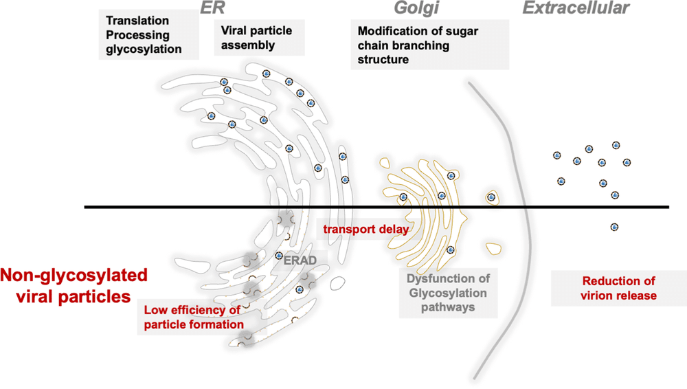 Fig.1
Model of the roles of prME N-glycosylation in viral particle formation and release.1
Fig.1
Model of the roles of prME N-glycosylation in viral particle formation and release.1
FAQs
Why is the glycosylation of the E protein variable between DENV and ZIKV?
While both viruses share the N153/N154 site, DENV possesses a second glycosylation site at N67. This N67 site is crucial for its specific interaction with DC-SIGN, influencing its tropism for dendritic cells. ZIKV lacks this N67 site, which may contribute to its differing tissue tropism and stability profile.
How does insect cell expression differ from mammalian expression for E protein studies?
Insect cells (vector host) typically produce paucimannose or high-mannose glycans, whereas mammalian cells (human host) produce complex, sialylated glycans. Since lectin binding (e.g., to DC-SIGN) is glycan-specific, using the correct expression system is vital for accurately modeling viral entry and pathogenesis.
What is the impact of glycosylation on vaccine design?
Glycosylation can mask neutralizing epitopes. A vaccine antigen with "incorrect" glycosylation might expose epitopes that are hidden on the native virus (leading to non-protective antibodies) or hide epitopes that should be targeted. Designing antigens with native-like glycosylation is crucial for eliciting a potent neutralizing response.
Can you analyze the glycan occupancy of my viral stock?
Yes. We offer mass spectrometry-based glycoprofiling services to determine both the site occupancy (macro-heterogeneity) and the specific glycan structures (micro-heterogeneity) present on your viral particles.
Does E protein glycosylation affect Antibody Dependent Enhancement (ADE)?
Yes. The glycan shield can modulate antibody binding affinity. Furthermore, the interplay between antibody binding and lectin receptor binding (mediated by glycans) can synergistically enhance viral uptake into Fc-gamma receptor-bearing cells, exacerbating ADE.
Reference:
- Ishida, K.; et al. N-linked glycosylation of flavivirus E protein contributes to viral particle formation. PLOS Pathogens. 2023, 19(10): e1011681. Distributed under Open Access license CC BY 4.0. https://doi.org/10.1371/journal.ppat.1011681