Anti-Keratan Sulfate (KS) Antibody Development Service
Keratan Sulfate (KS) represents one of the most structurally complex and biologically significant classes of sulfated glycosaminoglycans (GAGs). Predominantly localized in the extracellular matrix (ECM) of the cornea, cartilage, and central nervous system (CNS), these linear polymers of repeating disaccharide units play pivotal roles in maintaining tissue hydration, mediating cell signaling, and preserving structural integrity. Despite their critical importance in physiology and pathology, the development of high-affinity, high-specificity antibodies against KS remains a formidable scientific challenge. The inherent low immunogenicity of these carbohydrate antigens, combined with high structural homology across mammalian species, often results in poor antibody response and significant cross-reactivity.
At Creative Biolabs, we have established a premier platform dedicated to overcoming these obstacles. Leveraging decades of experience in carbohydrate immunology and advanced antibody engineering within our anti-glycosaminoglycan (GAG) antibody development platform, we offer a comprehensive service specifically for custom anti-keratan sulfate antibodies. Our proprietary technologies in glycan immunogen design and high-throughput screening allow us to generate reagents with exquisite specificity for defined sulfation motifs. We support researchers worldwide in elucidating the roles of KS in corneal dystrophy, skeletal disorders, inflammation, and tumor-associated glycan alterations, providing tools that are rigorously validated for research use.
Understanding Keratan Sulfate (KS) Biology
Keratan Sulfate is unique among GAGs as it lacks uronic acid. Instead, its backbone consists of repeating disaccharide units of galactose (Gal) and N-acetylglucosamine (GlcNAc), linked via alternating β(1→4) and β(1→3) glycosidic bonds. The structural diversity of KS arises primarily from the sulfation pattern at the C-6 position of either the Gal or GlcNAc residues, mediated by specific sulfotransferases such as CHST1 (KSGal6ST) and CHST6 (GlcNAc6ST). These sulfation modifications create highly anionic domains that interact with a wide array of growth factors, cytokines, and morphogens.
KS chains are covalently attached to core proteins to form proteoglycans. Based on the linkage type, KS is classified into three distinct subtypes:
- KS-I (Corneal Type): Linked via an N-glycosidic bond to asparagine residues on core proteins such as lumican, keratocan, and mimecan. KS-I is highly sulfated and essential for maintaining the spacing of collagen fibrils, which is requisite for corneal transparency.
- KS-II (Skeletal Type): Linked via an O-glycosidic bond to serine or threonine residues on cartilage proteoglycans like aggrecan. KS-II contributes to the compressive stiffness of cartilage and is a marker of tissue maturity and degeneration.
- KS-III (Brain Type): Linked via an O-glycosidic bond to mannose residues. Found extensively in the central nervous system on phosphacan and SV2, KS-III plays a dual role in neural plasticity and the inhibition of axonal regeneration following injury.
Challenges in Generating High-Affinity Anti-KS Antibodies
The production of reliable antibodies against KS epitopes is fraught with technical difficulties that standard antibody generation protocols often fail to address. Researchers frequently encounter the following bottlenecks:
Immunological Tolerance
As a highly conserved self-antigen, KS is poorly immunogenic in standard host animals. The mammalian immune system typically suppresses responses to these ubiquitous carbohydrate structures, leading to low titer and low affinity antibodies.
Structural Heterogeneity
KS chains are polydisperse and heterogeneous. A single proteoglycan may carry KS chains with varying lengths and sulfation degrees. Isolating an antibody that recognizes a specific "sulfation code" without binding to other motifs is extremely difficult.
Cross-Reactivity
Many GAG antibodies suffer from cross-reactivity with structurally related molecules such as Chondroitin Sulfate (CS), Dermatan Sulfate (DS), or Hyaluronic Acid (HA). This lack of specificity can lead to high background noise and misinterpreted experimental data.
Context Dependence
The presentation of the KS epitope can be influenced by the core protein. Antibodies generated against free glycan chains often fail to recognize the native epitope when it is embedded within the complex architecture of the ECM or cell surface.
Custom Anti-KS Antibody Development Services
To address these challenges, Creative Biolabs employs a multi-faceted approach. We utilize proprietary neoepitope immunogen design strategies, where short, defined KS oligosaccharides are conjugated to immunogenic carrier proteins (such as KLH or BSA) or displayed on virus-like particles (VLPs) to break immune tolerance. Furthermore, we offer distinct development tracks tailored to the specific isoform of interest.
Anti-KS-I (Corneal Type) Antibody Generation
We specialize in developing antibodies against the highly sulfated KS-I isoform predominant in the cornea. By using specific high-sulfated neoepitopes that mimic the structure found on lumican and keratocan, we generate high-affinity reagents critical for researching corneal transparency, wound healing, and Macular Corneal Dystrophy (MCD). These antibodies are validated to differentiate normal stromal tissue from pathological variants.
Anti-KS-II (Skeletal Type) Antibody Generation
Our platform offers targeted development for KS-II antibodies found in cartilage and bone matrices. By selecting immunogens that mimic the skeletal sulfation patterns associated with aggrecan, we produce antibodies essential for studying osteoarthritis, intervertebral disc degeneration, and Morquio syndrome. These tools are ideal for tracking cartilage turnover and matrix degradation in disease models.
Anti-KS-III (Brain Type) Antibody Screening
For the more elusive KS-III found in the central nervous system, which often exhibits lower sulfation and distinct mannose-linked structures, we employ our robust phage display technology. We screen massive synthetic and immune libraries against specific brain-derived KS glycan arrays to identify scFv or Fab binders. This high-throughput approach facilitates the discovery of rare clones that detect KS-III without cross-reacting with other CNS glycosaminoglycans.
Antibody Engineering and Labeling
Once a candidate anti-keratan sulfate antibody is identified—whether for KS-I, KS-II, or KS-III—we provide comprehensive downstream engineering services. This includes isotype switching to preferred formats, humanization for clinical research candidates, and direct conjugation. We can deliver your anti-KS antibody labeled with biotin, HRP, or fluorophores (e.g., FITC, PE, APC) for immediate use in flow cytometry or one-step tissue staining, streamlining your experimental workflow.
Project Workflow
Request a Quote for KS Antibody Service
Service Highlights
Defined Sulfation Specificity
Screening strategies designed to distinguish between high-sulfated and low-sulfated KS epitopes.
Strict Validation
Rigorous counter-screening against Chondroitin Sulfate and Dermatan Sulfate to prevent cross-reactivity.
Versatile Applications
Antibodies validated for ELISA, Immunohistochemistry (IHC), Immunofluorescence (IF), and Flow Cytometry.
Expert Consultation
PhD-level technical support to guide you from antigen selection to final assay development.
Applications of Anti-KS Antibodies
Corneal Research and Diagnostics
The transparency of the cornea depends heavily on the uniform arrangement of collagen fibrils, which is regulated by KS-proteoglycans like lumican and keratocan. In conditions such as Macular Corneal Dystrophy (MCD), a genetic defect prevents the proper sulfation of KS, leading to corneal opacification. Anti-KS antibodies are indispensable tools for:
- Studying the biosynthesis and sulfation pathways of corneal KS in normal and diseased states.
- Monitoring corneal wound healing and fibrosis where KS expression is typically downregulated or altered.
- Quality control of bioengineered corneal tissues for transplantation to ensure correct ECM composition.
Skeletal and Cartilage Disorders
In cartilage, KS is a major component of aggrecan, the large aggregating proteoglycan responsible for resisting compressive loads. Changes in serum or synovial fluid KS levels are potential biomarkers for osteoarthritis (OA) and rheumatoid arthritis (RA). Our antibodies enable researchers to:
- Quantify KS fragments in biological fluids as a specific marker of cartilage turnover and matrix degradation.
- Perform immunohistochemical analysis to map cartilage matrix degeneration in early-stage OA models.
- Investigate the pathogenesis of skeletal dysplasia, such as Mucopolysaccharidosis IV (Morquio A and B syndrome), where KS accumulation occurs.
Neuroscience and Regeneration
Following CNS injury, reactive astrocytes upregulate inhibitory proteoglycans in the glial scar. Highly sulfated KS has been identified as a potent inhibitor of neurite outgrowth and axonal regeneration. Researchers use our high-specificity anti-KS antibodies to:
- Precisely map the spatio-temporal distribution of inhibitory GAGs in spinal cord injury (SCI) and traumatic brain injury (TBI) models.
- Evaluate therapeutic strategies aimed at digesting the glial scar (e.g., Keratanase treatment) to promote regeneration.
- Study the role of KS in microglia activation, synaptic plasticity, and neuroinflammation.
Published Data
Understanding the diverse sulfation patterns of Keratan Sulfate (KS) in the central nervous system is crucial for developing specific antibodies. A recent review in the Journal of Neurochemistry highlights the complex organization of brain KS chains. The study illustrates how specific monoclonal antibodies recognize distinct sulfated regions within the KS chain structure.
As depicted in Figure 1, brain KS glycosaminoglycan chains are organized into specific di-sulfated and mono-sulfated regions. The figure demonstrates the binding sites for key monoclonal antibodies: clone 5D4 targets the highly sulfated (di-sulfated) heptasaccharide motifs, while clones like 1B4 and R10G recognize mono-sulfated regions. This structural insight underpins our strategy to develop custom antibodies that can selectively differentiate between these subtle sulfation patterns, providing researchers with precise tools to dissect KS function in neural development and repair.
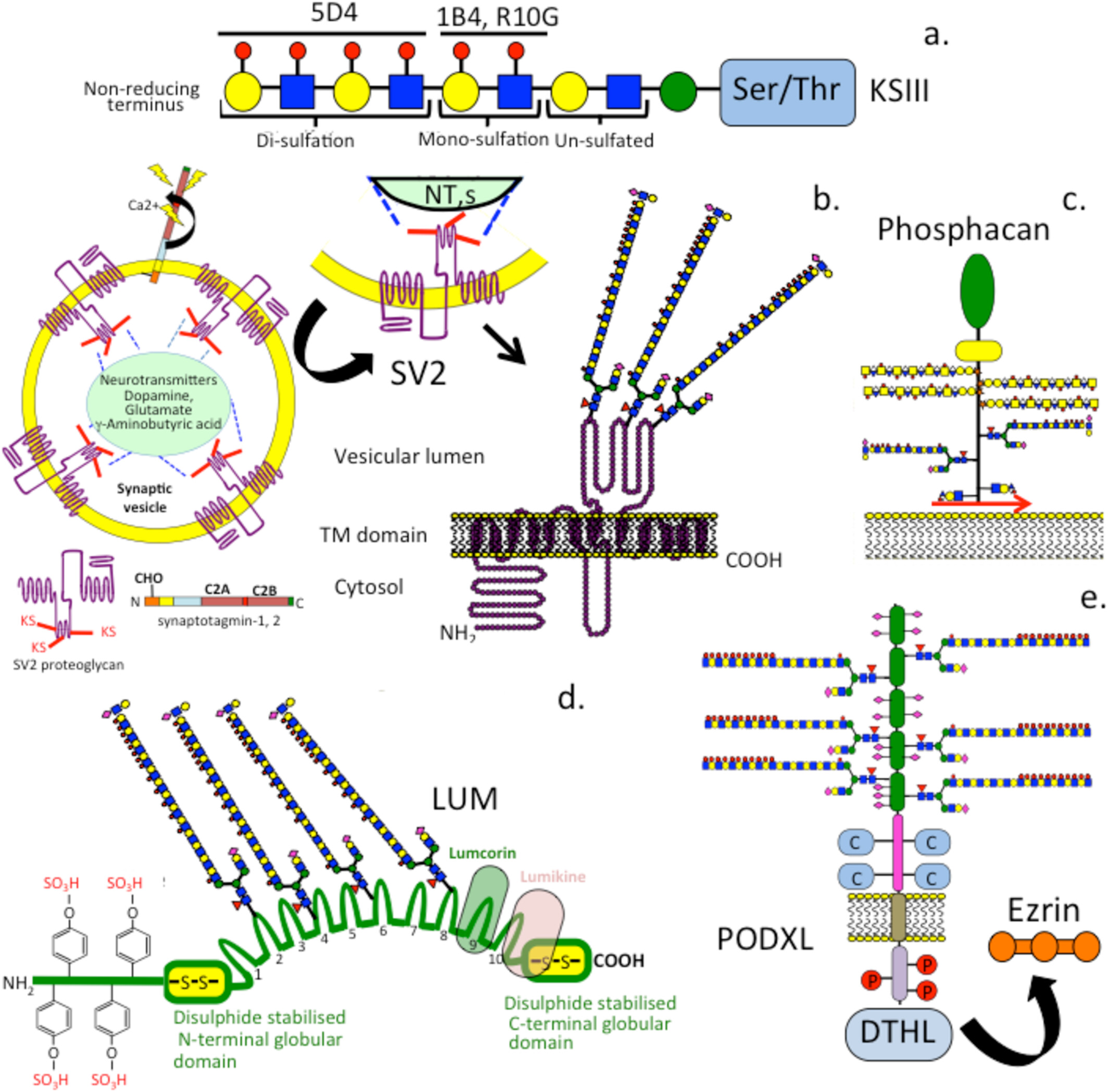 Fig.1 Organization of brain keratan sulfate (KS) chains and antibody recognition sites. Brain KS chains contain di- and mono-sulfated regions identified by specific monoclonal antibodies (5D4, 1B4, R10G).1
Reference:
- Melrose, James. "Keratan sulfate is a multifunctional brain glycosaminoglycan with instructive capabilities." Journal of Neurochemistry 169 (2025): e70208. Distributed under Open Access license CC BY 4.0. https://onlinelibrary.wiley.com/doi/10.1111/jnc.70208
FAQs
How do you ensure the antibody differentiates KS from Chondroitin Sulfate?
Cross-reactivity is a common issue in GAG antibody development. We implement a rigorous negative screening step. We screen our phage libraries or hybridoma supernatants against arrays containing Chondroitin Sulfate (CS), Dermatan Sulfate (DS), and Hyaluronic Acid (HA). Only clones that bind to KS but show no binding to these related GAGs are selected for further development.
Can you generate antibodies against specific sulfation patterns of KS?
Yes. KS chains vary in sulfation (e.g., non-sulfated, mono-sulfated, or di-sulfated disaccharides). By using specific oligosaccharide fragments or enzymatically modified KS as immunogens, we can direct the immune response toward specific sulfation motifs, such as high-sulfated KS found in the cornea versus low-sulfated forms in cartilage.
What applications are your anti-KS antibodies validated for?
Our standard validation package includes ELISA for affinity determination. Depending on your requirements, we can also validate the antibodies for Western Blot (using proteoglycan digests), Immunohistochemistry (IHC) on relevant tissue sections (e.g., cornea or cartilage), and Flow Cytometry.
Do you offer the 5D4 clone or can you make a similar antibody?
While we can produce recombinant versions of published sequences if freedom-to-operate allows, our primary expertise lies in developing novel custom antibodies. We can generate new clones with binding profiles similar to 5D4 (targeting high-sulfated KS) or clones with unique specificities that commercially available reagents do not cover.
Supports
- Anti-Heparan Sulfate (HS) Antibody Development Service
- Anti-Chondroitin Sulfate (CS) Antibody Development Service
- Anti-Dermatan Sulfate (DS) Antibody Development Service
- Anti-Keratan Sulfate (KS) Antibody Development Service
- Anti-Hyaluronic Acid (HA) Antibody Development Service
- Anti-GAG Sulfation Motif (Neo-epitope) Antibody Development Service
- Tumor-Associated GAG Antibody Development Service