HIV-1 Envelope Glycans: Targets for Broadly Neutralizing Antibodies
The Human Immunodeficiency Virus Type 1 (HIV-1) envelope glycoprotein (Env) is the sole viral antigen exposed on the virion surface, making it the primary target for neutralizing antibodies. However, the virus has evolved a sophisticated defense mechanism: a dense coating of host-derived carbohydrates known as the "glycan shield." This shield effectively masks conserved protein epitopes from immune recognition, allowing the virus to evade the host's humoral response. Despite this formidable defense, recent advances in structural biology and immunology have revealed that this shield is not impenetrable. At Creative Biolabs, we specialize in the complex biology of viral glycoproteins. Our advanced Anti-HIV Glycan Shield Antibody Development services are designed to help researchers exploit the vulnerabilities within the HIV envelope glycans, facilitating the discovery of next-generation broadly neutralizing antibodies (bNAbs) and the design of glycan-based vaccine immunogens.
The Architecture of the HIV-1 Glycan Shield
The functional HIV-1 envelope is a trimer of heterodimers, each consisting of a surface subunit (gp120) and a transmembrane subunit (gp41). What distinguishes Env from other viral glycoproteins is its exceptionally high glycosylation density. Approximately 50% of the molecular mass of the gp120 subunit consists of N-linked glycans. These glycans are attached to specific asparagine residues within the consensus sequence Asn-X-Ser/Thr (where X is any amino acid except proline).
The arrangement of these glycans creates a dynamic protective shell. Unlike the rigid protein core, the glycan shield is flexible and heterogeneous. This structural plasticity prevents the immune system from easily locking onto specific viral features. However, the density of glycosylation sites on Env is so high that it impedes the normal enzymatic processing of glycans in the host cell's endoplasmic reticulum and Golgi apparatus.
High-Mannose Glycans: A Structural Bottleneck
In typical mammalian glycoproteins, N-linked glycans are processed into "complex-type" structures capped with sialic acid. On the HIV-1 Env, specifically on the gp120 subunit, the crowding of glycans restricts the access of alpha-mannosidases, enzymes required for glycan maturation. This steric occlusion preserves a cluster of under-processed, oligomannose-type glycans (predominantly Man5-9GlcNAc2).
This cluster, often referred to as the "mannose patch," is distinct from the surrounding complex glycans and the "self" glycans found on healthy host cells. While the virus uses these host-derived molecules to mimic "self" and avoid immune detection, the unusual density and oligomannose composition create a unique antigenic surface that does not exist on normal human proteins. This inadvertent "non-self" feature has become a critical target for therapeutic intervention.
Mechanisms of Immune Evasion
The HIV-1 glycan shield employs several strategies to thwart antibody neutralization:
- Steric Hindrance: Large glycan trees physically block antibodies from reaching the conserved protein epitopes, such as the CD4 binding site (CD4bs).
- Molecular Mimicry: Because the glycans are synthesized by the host cell machinery, they are chemically identical to host glycans, reducing their immunogenicity.
- Shifting Landscape: The virus can rapidly mutate the location of N-linked glycosylation sites (PNGS), effectively moving the holes in the shield and rendering strain-specific antibodies useless.
Targeting Glycans with Broadly Neutralizing Antibodies (bNAbs)
Despite these defenses, a subset of HIV-1 infected individuals eventually develops broadly neutralizing antibodies (bNAbs) that can traverse or directly recognize the glycan shield. These antibodies are categorized based on their epitopes, and several key classes target the gp120 glycosylation specifically.
The PGT Family (PGT121, PGT128, PGT135)
These highly potent bNAbs target the V3 loop base and its surrounding glycans. For example, PGT128 inserts its long HCDR3 loop into the glycan shield, contacting both the peptide backbone and the specific high-mannose glycans at position N332. This "glycan-dependent" recognition allows the antibody to anchor itself securely to the virus.
The 2G12 Antibody
2G12 is unique among bNAbs because it binds exclusively to carbohydrates. It possesses an unusual "domain-swapped" structure that creates a multivalent binding surface capable of recognizing the dense cluster of Man9GlcNAc2 glycans on the outer domain of gp120. It demonstrates that the glycan shield itself can be a target, not just a barrier.
V1/V2 Glycan-Dependent bNAbs (PG9, PG16)
This class of antibodies targets the apex of the Env trimer. They recognize a quaternary epitope formed by the V1/V2 loops and their associated glycans (specifically N160). Binding often depends on the specific sialylation state of the glycans, highlighting the importance of precise glycosylation analysis in immunogen design.
Interface bNAbs (8ANC195)
Some antibodies target the gp120/gp41 interface, often incorporating glycans like N234 and N276 into their epitope footprint. These bNAbs stabilize the trimer in a non-functional state and prevent the conformational changes required for viral fusion.
Comprehensive Services for HIV Glycan Research
The development of antibodies against the HIV glycan shield requires specialized tools and expertise. Standard immunization protocols often fail to generate responses against these low-immunogenicity carbohydrate targets. Creative Biolabs provides an integrated suite of services to overcome these challenges.
Anti-HIV Glycan Shield Antibody Development
We offer custom antibody generation strategies tailored for the HIV envelope. This includes the design of native-like Env trimers, glycan-engineered immunogens, and specialized B-cell sorting protocols to isolate rare clones that recognize glycan-dependent epitopes.
Glycoarray Platforms
Our high-throughput glycan arrays allow for the rapid profiling of antibody specificity. We can screen your candidate bNAbs against diverse panels of high-mannose, complex, and hybrid glycans to map their precise binding requirements.
Glycosylation Analysis
Understanding the exact composition of your immunogen is critical. We provide site-specific glycosylation analysis using mass spectrometry to verify that your recombinant Env proteins display the correct "mannose patch" architecture required for bNAb elicitation.
Custom Glycosylation of Biomolecules
We can enzymatically or chemically modify the glycan profile of your HIV antigens. Whether you need to enrich for high-mannose structures or remove specific decoys, our remodeling services ensure your research materials are optimized for study.
Inquire About HIV Glycan Services
Published Data
Recent computational analyses utilizing microsecond-scale molecular dynamics simulations have provided an atomistic view of the HIV-1 envelope glycan shield, revealing it as a dynamic and interconnected network rather than a static coating. This research quantified the solvent-accessible surface area of the trimer, demonstrating that while the glycan canopy covers approximately 70% of the protein surface, it is maintained by specific inter-glycan interactions that limit the conformational freedom of individual sugar moieties.
Crucially, the study mapped the existence of persistent "glycan holes"—regions of vulnerability where the protein surface remains exposed due to steric constraints that prevent complete glycan coverage. These vulnerabilities, particularly near the CD4 binding site and the trimer apex, align precisely with the epitopes of known broadly neutralizing antibodies. The data further revealed that the processing state of specific glycans (high-mannose versus complex) dictates the integrity of these networks; the removal or modification of a single glycan can destabilize local shielding, creating new avenues for immune recognition. These findings underscore the necessity of replicating native-like glycosylation patterns in vaccine immunogens to ensure that these neutralizing epitopes are presented in their physiologically relevant, accessible state, rather than being inadvertently occluded by non-native glycan clustering.
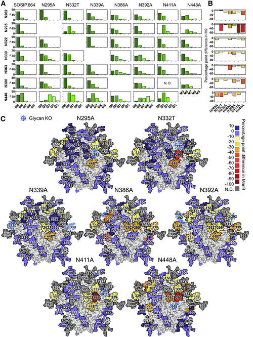 Fig.1
Molecular Dynamics Visualization of the HIV-1 Glycan Shield Density and Epitope Accessibility.1
Fig.1
Molecular Dynamics Visualization of the HIV-1 Glycan Shield Density and Epitope Accessibility.1
FAQs
Why are high-mannose glycans so prevalent on HIV-1 gp120?
The unusually high density of glycosylation sites on gp120 creates steric crowding. This crowding prevents alpha-mannosidase enzymes in the Golgi from accessing the glycans to process them into complex structures. As a result, a cluster of under-processed, oligomannose (high-mannose) glycans remains, known as the "mannose patch."
Can antibodies be developed that target only the glycans?
Yes, the most famous example is 2G12, which binds exclusively to the mannose patch on gp120 without contacting the protein backbone. However, most glycan-reactive bNAbs (like the PGT family) target a mixed epitope consisting of both specific glycans and the underlying peptide sequence.
What is the biggest challenge in developing vaccines based on the glycan shield?
The primary challenge is heterogeneity. The glycans on the viral surface are structurally diverse and flexible ("wobbly"). A successful vaccine immunogen must stabilize these glycans in a specific conformation that mimics the native viral spike to trigger the production of broadly neutralizing antibodies rather than non-neutralizing binding antibodies.
Does Creative Biolabs provide native-like HIV-1 Env trimers?
Yes. We utilize advanced expression systems (such as CHO or 293T cells) and purification techniques (like SOSIP stabilization) to produce soluble Env trimers that closely mimic the native viral spike, preserving the quaternary structure and the authentic glycan shield profile.
How do you validate the glycan binding specificity of an antibody?
We use a combination of techniques. Glycan microarrays allow us to screen binding against hundreds of defined carbohydrate structures. We also employ site-directed mutagenesis (removing specific glycosylation sites) and enzymatic deglycosylation assays to confirm if antibody binding is glycan-dependent.
Reference:
- Seabright, G.E., et al. "Networks of HIV-1 Envelope Glycans Mask and Shield Neutralizing Epitopes in Silico." Structure 28.8 (2020): 897-909. Distributed under Open Access license CC BY 4.0, without modification. https://doi.org/10.1016/j.str.2020.04.022