Influenza Virus Hemagglutinin and Neuraminidase Glycosylation
Influenza A viruses pose a persistent threat to global health, largely due to the rapid evolution of their surface glycoproteins. The intricate interaction between the host immune system and the viral surface is mediated primarily by hemagglutinin (HA) and neuraminidase (NA). These proteins are heavily glycosylated, and their glycosylation patterns are not static; they evolve dynamically to shield the virus from antibody neutralization. At Creative Biolabs, we provide specialized solutions for characterizing these complex structures, including our advanced Anti-Influenza Virus Glycan Shield Antibody Development service. Understanding the structural biology of HA and NA glycosylation is critical for developing next-generation therapeutics and broader-spectrum vaccines.
Structural Biology of Influenza Surface Glycoproteins
The surface of the influenza virus is studded with two primary glycoproteins that determine its antigenicity and host specificity: hemagglutinin and neuraminidase. Both proteins utilize the host cell's post-translational modification machinery to attach complex oligosaccharides to specific amino acid residues. These glycans are essential for protein folding, stability, and transport, but they also serve a crucial role in immune evasion by creating a "glycan shield" that masks antigenic sites.
Hemagglutinin (HA) Glycosylation
HA is a trimeric glycoprotein responsible for binding to sialic acid receptors on the host cell surface and mediating viral entry. It is synthesized as a precursor (HA0) which is cleaved into two subunits: the globular head (HA1) and the stem (HA2). The influenza hemagglutinin glycosylation sites are primarily N-linked, attaching to asparagine residues within the consensus motif Asn-X-Ser/Thr.
The glycans on the HA stem are generally conserved across different strains because they are structurally vital for the trimer's stability. However, the globular head, which contains the receptor-binding site (RBS) and the major antigenic epitopes, exhibits high variability in glycosylation. During viral evolution, the acquisition of new glycosylation sites on the HA head physically sterically hinders antibody access, preventing neutralization without compromising receptor binding.
Neuraminidase (NA) Structure
NA is a tetrameric enzyme that cleaves terminal sialic acid residues from cell surface glycoconjugates. This enzymatic activity is essential for preventing viral aggregation and facilitating the release of progeny virions from the infected cell. Like HA, neuraminidase structure is heavily influenced by N-linked glycosylation.
Glycans on NA are critical for its enzymatic stability and tetramer formation. Interestingly, the balance between HA binding affinity and NA cleavage activity is delicate; changes in glycosylation on one protein often drive compensatory mutations in the other. Recent studies suggest that glycosylation on the NA head domain can also mask epitopes, contributing to the virus's ability to escape inhibition by NA-specific antibodies, which are increasingly recognized as independent correlates of protection.
Comparative Glycosylation Profiles of Influenza A Subtypes
To understand the distinct evolutionary pressures and structural constraints acting on different viral domains, it is essential to compare how glycosylation sites are distributed and conserved across the viral surface proteins. The table below summarizes these key differences between the HA and NA domains.
| Viral Protein Domain | Glycosylation Characteristics | Evolutionary Trend | Functional Impact |
|---|---|---|---|
| HA Globular Head | Highly variable N-linked sites | Rapid accumulation (e.g., H3N2) to mask epitopes | Immune evasion; modulation of receptor affinity |
| HA Stem | Highly conserved N-linked sites | Stable across subtypes | Trimer stability; fusion peptide regulation |
| NA Head | Variable; typically 3-5 sites | Site shifting to cover active site perimeter | Enzymatic activity modulation; tetramer stability |
| NA Stalk | Variable length and glycosylation | Deletions often observed in adaptation | Determines viral morphology and access to substrate |
Multifaceted Roles of Glycans in Viral Life Cycle
Beyond passive shielding, N-linked glycans actively regulate critical steps in the influenza life cycle:
- Proteolytic Activation: Glycans located near the cleavage site of the HA0 precursor can modulate accessibility to host proteases, a rate-limiting step for viral infectivity.
- Receptor Specificity Tuning: Glycans adjacent to the Receptor Binding Site (RBS) can sterically hinder binding to sialic acid receptors, often necessitating compensatory mutations to maintain fitness during host adaptation.
- Virion Release Balance: Glycosylation on Neuraminidase (NA) affects its enzymatic efficiency. An optimal balance between HA binding and NA cleavage is maintained through co-evolution of glycosylation sites on both proteins.
- Antigenic Decoys: Some glycans can mimic host structures, reducing the immunogenicity of the virus by exploiting the host's tolerance mechanisms.
Mechanisms of Antigenic Drift via Glycosylation
Antigenic drift is the accumulation of mutations that allow the virus to escape pre-existing immunity. While amino acid substitutions are a primary driver, the alteration of glycosylation motifs is a distinct and powerful mechanism of flu virus evolution.
The "Glycan Shield" Phenomenon
When the human population develops high levels of immunity against a circulating strain, the virus is under immense selective pressure. Variants that acquire mutations introducing a new glycosylation site (e.g., an N-linked sequon) on the HA head can survive because the bulky glycan physically blocks antibodies from binding to the underlying protein epitope. This is often referred to as "epitope masking."
Over decades, H3N2 subtypes have shown a progressive accumulation of glycans on the HA head. While this aids in immune evasion, it can come at a fitness cost by interfering with receptor binding. The virus often compensates with secondary mutations to restore affinity, illustrating the complex evolutionary trade-offs involved in maintaining the glycan shield.
Host Receptor Binding and Species Specificity
The host receptor binding specificity of influenza is governed by the linkage of sialic acids: human viruses prefer alpha-2,6 linkages, while avian viruses prefer alpha-2,3 linkages. Glycosylation near the receptor-binding pocket can modulate this specificity.
For a virus to cross the species barrier (zoonosis), changes in glycosylation are often required to unmask the receptor-binding site or alter its geometry to accommodate human-type receptors. Therefore, monitoring glycosylation patterns is not only important for vaccine strain selection but also for pandemic risk assessment.
Advanced Services for Influenza Glycan Research
Creative Biolabs offers a comprehensive suite of services designed to dissect the complexity of viral glycoproteins. From developing antibodies that target cryptic epitopes to mapping the glycan shield, our platform supports virologists and vaccine developers worldwide.
Anti-Influenza Virus Glycan Shield Antibody Development
Targeting the glycan shield requires specialized strategies. We develop broadly neutralizing antibodies (bnAbs) that can penetrate the glycan density or recognize the glycan-protein interface. Our service includes phage display library screening and B-cell sorting strategies optimized for highly glycosylated viral antigens.
Glycoarray Platforms
To determine the receptor specificity of your viral isolates, we offer high-throughput glycoarray screening. Our arrays feature a diverse library of sialylated glycans, including human (alpha-2,6) and avian (alpha-2,3) structures, allowing for rapid profiling of HA binding preferences and potential host range shifts.
Glycosylation Analysis
We provide detailed structural characterization of viral glycoproteins using mass spectrometry (LC-MS/MS). We can map N-linked glycosylation sites, determine site occupancy, and analyze the composition of the attached glycans (high mannose, hybrid, or complex) to validate your vaccine antigens or recombinant proteins.
Custom Glycosylation of Biomolecules
For research requiring specific glycoforms, we offer custom glycoprotein engineering. We can express HA and NA antigens in mammalian, insect, or yeast systems with tailored glycosylation profiles to study the impact of specific glycans on immunogenicity and antibody binding.
Inquire About Glycan Analysis Services
Published Data
A pivotal study published in PLOS Pathogens elucidates the intricate evolutionary trade-offs influenza A viruses navigate when escaping neutralizing antibodies. While antigenic escape substitutions are essential for evading host immunity, they frequently impose significant fitness costs by disrupting the optimal geometry of the Hemagglutinin (HA) receptor-binding pocket, leading to reduced cell entry efficiency. The research highlights a sophisticated compensatory mechanism wherein the virus rapidly selects for de novo N-linked glycosylation sites on the HA globular head to recover lost fitness.
Challenging the conventional paradigm that viral glycans function solely as steric shields against antibody recognition, this investigation reveals their structural role in allosteric modulation. Using advanced kinetic analysis, researchers demonstrated that specific glycan additions—emerging naturally after antibody pressure was removed—successfully restored high-affinity interactions with sialic acid receptors. As shown in Fig.1, Bio-layer interferometry (BLI) data confirms that the introduction of a glycan at the rim of the receptor-binding site (e.g., mutant N133T) corrects the biophysical defects caused by prior immune escape mutations, effectively re-tuning the virus's binding avidity to near wild-type levels. This data underscores the multifaceted nature of the "glycan shield," demonstrating that it serves a dual purpose: fortifying immune evasion while simultaneously stabilizing essential protein functions.
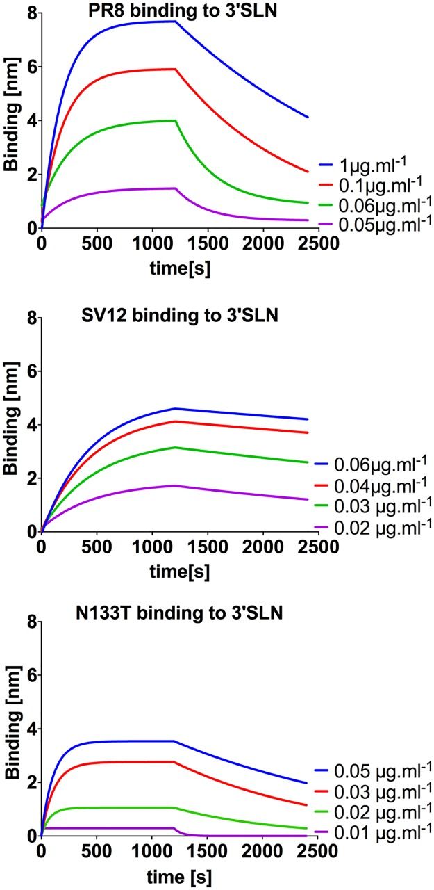 Fig.1
Kinetic analysis revealing the compensatory role of HA glycosylation in restoring receptor-binding avidity.1
Fig.1
Kinetic analysis revealing the compensatory role of HA glycosylation in restoring receptor-binding avidity.1
FAQs
How does HA glycosylation contribute to influenza immune evasion?
HA glycosylation contributes to immune evasion by creating a "glycan shield." These oligosaccharides are bulky and can physically cover (mask) conserved antigenic epitopes on the HA surface. Since the glycans are derived from the host, the immune system often recognizes them as "self" and does not produce antibodies against them, effectively hiding the virus from neutralizing antibodies.
What is the difference between antigenic drift and antigenic shift?
Antigenic drift refers to the gradual accumulation of small mutations (amino acid substitutions or glycosylation changes) in HA and NA, requiring annual vaccine updates. Antigenic shift is a sudden, major change resulting from the reassortment of gene segments between different influenza viruses (e.g., avian and human), potentially causing pandemics.
Why is the N-X-S/T motif important in influenza evolution?
The Asn-X-Ser/Thr (N-X-S/T) motif is the consensus sequence required for N-linked glycosylation. Influenza viruses frequently evolve by acquiring mutations that create this motif on the HA head, allowing the attachment of a new glycan to mask an antibody binding site.
Can Creative Biolabs analyze the specific glycan structures on my viral isolate?
Yes. We utilize advanced liquid chromatography-tandem mass spectrometry (LC-MS/MS) and glycoarray platforms to precisely map glycosylation sites and identify the specific glycan structures (e.g., high mannose vs. complex) present on HA and NA proteins from your specific viral isolate.
Does neuraminidase (NA) glycosylation affect viral fitness?
Yes. NA glycosylation is crucial for the stability and tetramerization of the enzyme. Changes in NA glycosylation can alter its enzymatic activity (cleavage of sialic acids), which must be balanced with HA binding affinity for optimal viral replication and transmission.
Reference:
- Kosik, I., et al. "Influenza A virus hemagglutinin glycosylation compensates for antibody escape fitness costs." PLOS Pathogens 14.1 (2018): e1006796. Distributed under Open Access license CC BY 4.0. https://doi.org/10.1371/journal.ppat.1006796
Supports
- Glycoarray Platforms
- Glycosylation Analysis
- Custom Glycosylation of Biomolecules
- Anti-Influenza Virus Glycan Shield Antibody Development