Lewis Antigen Introduction
Accelerate Your Glycan-Based Discoveries!
Are you currently facing challenges in identifying specific glycan biomarkers, grappling with complex glycosylation pattern analysis, or navigating long development cycles in glycan-related drug discovery? Creative Biolabs' comprehensive Lewis Antigen solutions help you accelerate glycan-related drug discovery, obtain precise glycan biomarker identification, and streamline glycosylation analysis through advanced glycoengineering, high-throughput glycan profiling, and innovative antibody development techniques.
Contact our team to get an inquiry now!
Lewis Antigen
Lewis (Le) antigens comprise fucose-modified saccharides (oligosaccharides) conjugated with lipids or proteins, thereby occurring as glycolipids or glycoproteins. Lewis antigens function as GPCRs and are chiefly produced by endodermal epithelia, though detected in these tissues and erythrocytes following glycolipid transfer to RBCs. Lewis expression in healthy tissues occurs on two principal saccharide chains: type 1 and type 2, distinguished by Gal-GlcNAc linkage (β1,3 versus β1,4). Type 1 chains contain Lewisa/b (Le-a/b), while type 2 chains express Lewisx/y (Le-x/y).
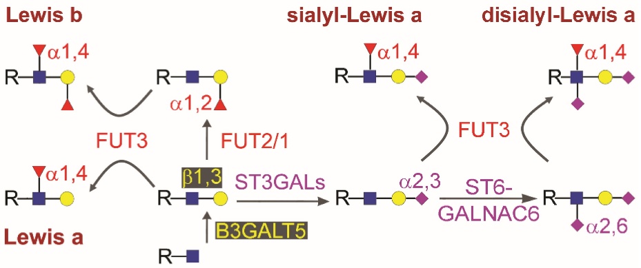 Fig.1 Processing and presentation of diverse carbohydrate antigen structures.1,4
Lewisa and Lewisb represent the principal antigens within the Lewis blood group system. Lewisb synthesis requires two fucosyltransferases (FUTs): the Lewis enzyme (Fuc-TIII; FUT3) encoded by the Lewis gene (an α(1,3/1,4)fucosyltransferase) and an α(1,2)fucosyltransferase, the latter being unnecessary for Lewisa production. Three prevalent phenotypes exist: Le(a+b-), Le(a-b+), and Le(a-b-). Naturally formed Lewis antibodies, predominantly IgM class, occur solely in Le(a-b-) individuals. Lewis antigens demonstrate comparable functions in healthy adult tissues, though context-dependent. Lewisx expression is observed in colon, stomach, salivary glands, kidneys, bladder, uterus, cervix, epididymis, and medulla epithelia, whereas Lewisy appears chiefly in breast, lung, colon, stomach, pancreas, uterus, ovary, prostate, salivary gland epithelia, and small intestinal Paneth cells.
Biosynthesis
The Lewis locus encodes fucosyltransferases (FUTs) that generate Lewis antigens. These enzymes exhibit expression patterns analogous to secretor loci.
Lewis enzyme FUT3 transfers fucose from GDP-Fuc to GlcNAc within type 1/2 chains. Attachment to type 2 chains yields an α3 linkage, while transfer to type 1 chains produces an α4 linkage, dictated by prior Gal occupancy at GlcNAc's 4 or 3 position respectively. This fucose addition creates Lewisa or Lewisx. α1-2-fucosylation of terminal Gal forms H antigen before α3/4FucT action, enabling Lewisb/y formation. H antigen synthesis employs identical α1-2FucT used for H precursor generation in ABO blood groups. In summary, Lewis antigens form via α3/4-fucose attachment: to unsubstituted type 1/2 chains yielding Lewisa/x, or to H type 1/2 chains producing Lewisb/y.
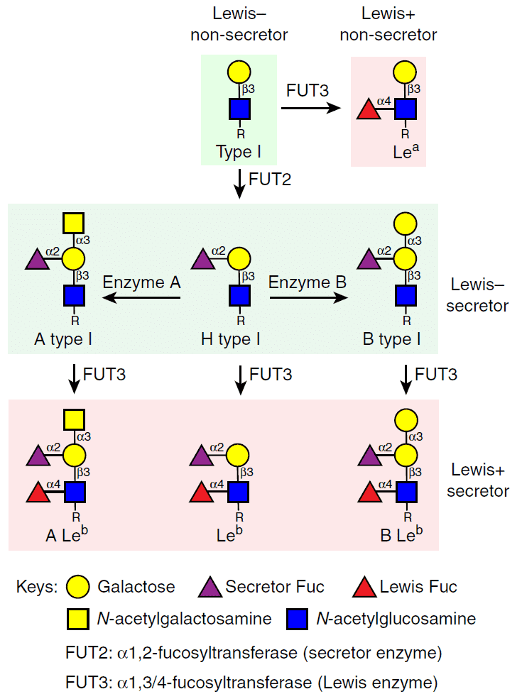 Fig.2 Synthetic pathway for type I histo-blood group antigens in secretor/non-secretor individuals.2,4
Functions & Clinical Values
Functionally, Lewisx mediates neutrophil transepithelial migration and delivers stimulatory immunomodulation to dendritic cells via engagement of the C-type lectin receptor DC-SIGN. Concurrently, Lewisx serves as the primary fucosylated brain antigen, mediating cell-cell interactions critical for neurodevelopment. It constitutes the stage-specific embryonic antigen-1 (SSEA-1) epitope and functions as a surface biomarker for neural stem cell identification.
Aberrant glycosylation represents a universal tumorigenesis hallmark. Fucosylated epitopes—including type I (H1, Lewisa, Lewisb, sialyl Lewisa) and type II (H2, Lewisx, Lewisy, sialyl Lewisx) Lewis antigens—are frequently overexpressed on malignant cells, principally due to upregulated relevant FUT expression. However, cancer cell impacts of fucosylated moieties are not fully understood.
Published Data
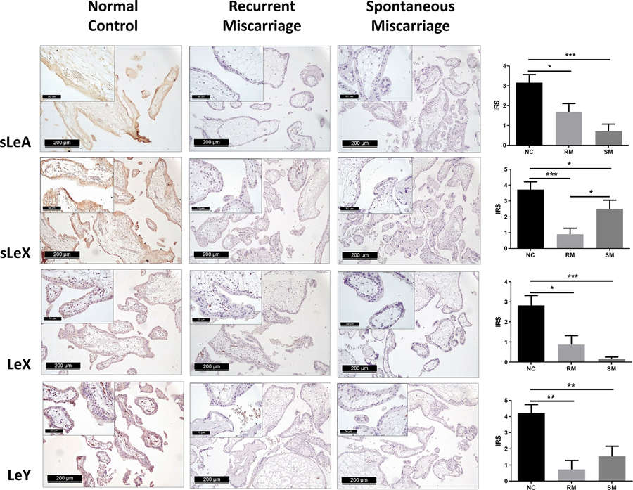 Fig.3 Immunohistochemical detection of four Lewis carbohydrate antigens in syncytiotrophoblasts across three placental tissue groups.3,4
Recent investigations into Lewis antigen expression patterns have provided compelling insights into their role in complex biological processes beyond oncology. One study examined the expression of Sialyl Lewis A (sLeA), Sialyl Lewis X (sLeX), Lewis X (LeX), and Lewis Y (LeY) in the placental villi tissue of patients with a history of unexplained miscarriages, comparing them to a control group with normal pregnancies.
The experimental findings demonstrated a significant upregulation of sLeA, sLeX, LeX, and LeY in the syncytiotrophoblast of the control group when contrasted with both spontaneous and recurrent miscarriage groups. While no substantial differences were observed between the spontaneous and recurrent miscarriage groups, the study identified interesting modulations in key enzymes: ST3GAL6 was significantly downregulated in the recurrent miscarriage group, whereas NEU1 was upregulated in the spontaneous group. Furthermore, LeX and LeY were observed in villous vascular endothelial cells of controls but became undetectable in spontaneous abortion groups. Simultaneous evaluation showed markedly reduced villous vessel density (via CD31 staining) across all placental villi size categories in cases of pregnancy loss. These findings collectively demonstrate a unique placental Lewis antigen distribution pattern in miscarriage versus normal gestation, suggesting novel mechanistic roles in its pathogenesis.
What We Can Offer?
Creative Biolabs is your trusted partner for advanced Lewis Antigen research and development. We provide a comprehensive range of products and services designed to meet the diverse and evolving needs of academic and industrial clients seeking to unravel the complexities of glycans in health and disease:
- Lewis Antigen Profiling & Quantification
- Anti-Lewis Antigen Antibody Development
- Functional Characterization of Lewis Antigens
- Lewis Antigen-Based Biomarker Discovery & Validation
- Custom Lewis Glycan Array Services
Leverage the Creative Biolabs Advantage – Obtain Your Quote Today
Why Choose Us?
Selecting Creative Biolabs for Lewis Antigen studies delivers distinct benefits, grounded in profound knowledge, advanced technologies, and steadfast dedication to customer achievement.
- Unrivaled Glycomics Expertise
- State-of-the-Art Analytical Platforms
- Customized & Flexible Solutions
- Robust Data Quality & Reproducibility
FAQs
Q: How can glycan analysis, specifically of Lewis Antigens, contribute to early cancer detection strategies?
A: While certain Lewis antigens, like CA19.9 and Lewisa/b, are widely recognized as valuable cancer markers for disease management, their elevated serum levels often manifest at later disease stages. This makes them generally less suitable for broad population-level early cancer screening in asymptomatic individuals. However, detailed Lewis antigen profiling can be exceptionally crucial for monitoring disease progression in diagnosed patients, assessing their response to various therapies, and predicting disease recurrence.
Q: What types of biological samples are typically suitable for Lewis Antigen analysis, and what sample preparation considerations are involved?
A: High-resolution Lewis Antigen analysis can be performed on a wide array of biological samples. These commonly include various human or animal cell lines (e.g., cancer cell lines, immune cells), both fresh and formalin-fixed paraffin-embedded (FFPE) tissue samples (such as tumor biopsies or adjacent normal tissue controls), and diverse biological fluids like serum, plasma, urine, and cerebrospinal fluid. Comprehensive sample preparation, including precise glycan release and purification, is a critical step to ensure optimal integrity and yield for subsequent analysis.
Q: What measures are taken to ensure the high specificity of antibodies developed against Lewis Antigens?
A: The development of highly specific antibodies targeting Lewis Antigens is a paramount objective. This involves a rigorous, multi-stage approach. Key measures include sophisticated antigen design to present the desired epitopes effectively, advanced immunization strategies, stringent and multi-layered screening protocols to identify high-affinity binders, and meticulous antibody engineering techniques.
Related Products and Services
To further advance your glycobiology R&D, we provide a portfolio of products:
- Monoclonal Antibodies
- Polyclonal Antibodies
- Secondary & Tag Antibodies
- Isotype & Loading Control Antibodies
- Carbohydrate Antigens
Creative Biolabs provides tailored anti-glycan antibody services for international clientele.
To explore these capabilities, please contact us for more information.
References:
- Indellicato, Rossella et al. "Complementary Use of Carbohydrate Antigens Lewis a, Lewis b, and Sialyl-Lewis a (CA19.9 Epitope) in Gastrointestinal Cancers: Biological Rationale Towards A Personalized Clinical Application." Cancers vol. 12,6 1509. 9 Jun. 2020, DOI:10.3390/cancers12061509
- Hu, Liya et al. "Glycan recognition in globally dominant human rotaviruses." Nature communications vol. 9,1 2631. 6 Jul. 2018, DOI:10.1038/s41467-018-05098-4
- Ma, Zhi et al. "Expression of the Carbohydrate Lewis Antigen, Sialyl Lewis A, Sialyl Lewis X, Lewis X, and Lewis Y in the Placental Villi of Patients With Unexplained Miscarriages." Frontiers in immunology vol. 12 679424. 31 May. 2021, DOI:10.3389/fimmu.2021.679424
- Distributed under Open Access license CC BY 4.0, without modification.