Custom Fluorescent Lipid Synthesis Service
Background Our Services Workflow Applications Why Choose Us Related Services FAQs
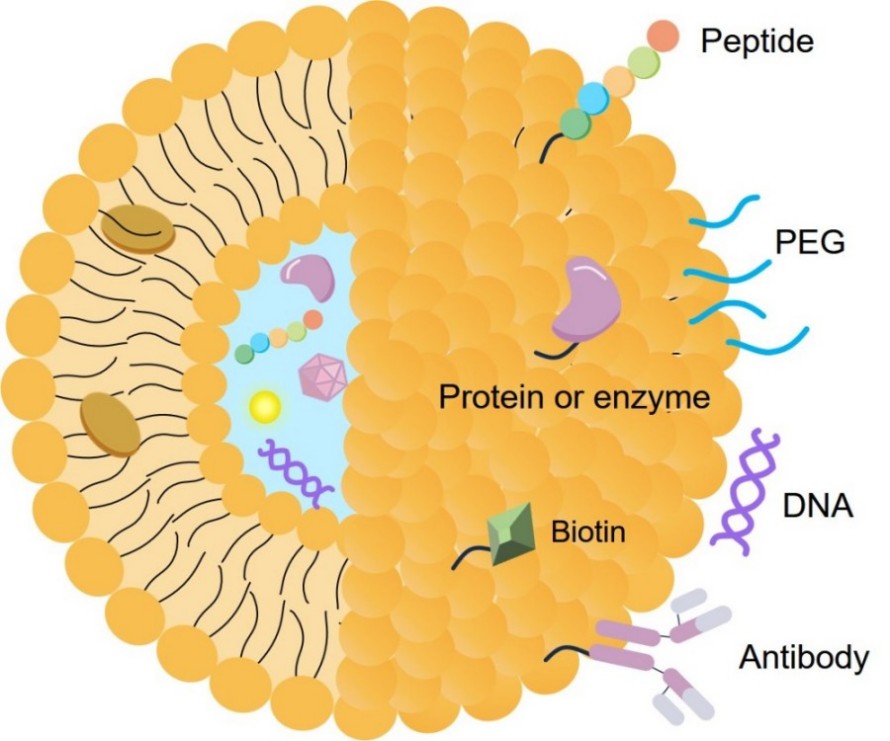
In the precise world of lipid-based drug delivery, the ability to visualize the journey of a carrier is as vital as the therapeutic payload itself. From tracking endosomal escape in mRNA vaccines to mapping the tissue biodistribution of novel liposomes, high-fidelity imaging is the key to optimization. However, standard off-the-shelf probes often lack the specificity or structural compatibility required for advanced formulations. At Creative Biolabs, we bridge this gap by offering expert Fluorescent Lipids Synthesis Services, creating high-purity, biomimetic probes that allow you to visualize lipid dynamics without compromising the integrity of your delivery system.
Fundamental Principles of Fluorescent Lipids
To effectively utilize fluorescent lipids in advanced research, it is essential to understand the intricate balance between signal detection and molecular mimicry. A fluorescent lipid is not simply a dye attached to a fat molecule; it is a carefully engineered tool designed to report on its environment without disturbing it.
The "Observer Effect" in Lipid Imaging
Just as in physics, the act of observing (labeling) a biological system can alter its behavior. A poorly designed probe acts as an impurity within the membrane, potentially causing:
-
Membrane Instability: Lowering the phase transition temperature (Tm), causing the liposome to leak prematurely.
-
The "Looping" Effect: If a hydrophilic fluorophore is attached to a hydrophobic tail, it may "snorkel" back to the water interface, bending the lipid chain and creating membrane defects.
-
Artifactual Distribution: The probe may segregate into specific domains not because of the lipid type, but because of the dye's preference, leading to false conclusions about lipid rafts.
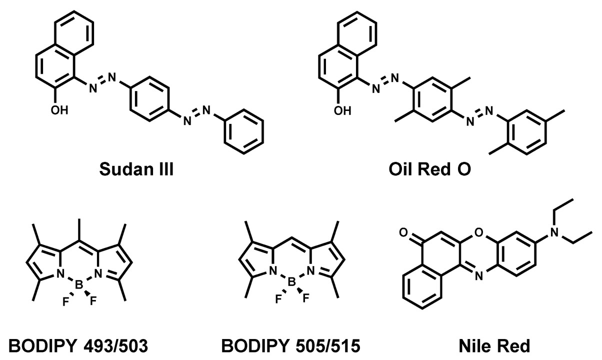 Fig. 1 Chemical structures of commonly used fluorescent dyes.1
Comparison of Common Fluorophores
Selecting the right fluorophore is a critical decision that depends on your specific imaging equipment and biological question. Below is a comparison of standard fluorophores used in lipid conjugation:
|
Fluorophore Family
|
Emission Color
|
Key Characteristics
|
Application
|
|
NBD
|
Green
|
Fluorescence intensity increases significantly in non-polar (membrane) environments.
|
Membrane fusion assays, lipid exchange studies.
|
|
Rhodamine (Lissamine)
|
Red
|
Excellent photostability and distinct spectral separation from GFP/FITC.
|
General membrane labeling.
|
|
Cyanine (Cy5, Cy7)
|
Far-Red/NIR
|
Near-Infrared emission avoids tissue autofluorescence.
|
In vivo biodistribution, animal imaging.
|
|
Pyrene
|
UV/Blue
|
Forms dimers at high concentrations, shifting emission wavelength.
|
Measuring membrane fluidity and lateral diffusion rates.
|
Optimization of Fluorophore Positioning
The functional efficacy and biophysical fidelity of a fluorescent lipid are intrinsically dictated by the specific conjugation site of the fluorophore. To ensure optimal compatibility with diverse experimental paradigms, we employ two distinct structural modification strategies:
Head-Group Labeling (Hydrophilic Modification)
-
Mechanism: The fluorophore is covalently attached to the polar headgroup (e.g., the primary amine of Phosphatidylethanolamine, PE).
-
Advantages: Maintains the integrity of the hydrophobic acyl chains, preserving the native packing parameter and bilayer thickness.
-
Considerations: The presence of a bulky fluorophore at the lipid-water interface may introduce steric hindrance, potentially modulating ligand-receptor kinetics or altering endocytic uptake mechanisms.
Acyl Chain Labeling (Hydrophobic Modification)
-
Mechanism: The fluorophore is integrated into the fatty acid tail, typically substituting a segment of the sn-2 acyl chain via precise organic synthesis.
-
Advantages: Sequesters the reporter within the hydrophobic bilayer core, significantly reducing spontaneous inter-membrane transfer (flip-flop) and eliminating surface steric artifacts.
-
Considerations: Synthetic complexity is higher; precise engineering of the linker and chain length is required to minimize perturbation of the membrane's Tm.
Comprehensive Custom Synthesis Solutions
Creative Biolabs provides a comprehensive, modular platform for the custom synthesis of fluorescently labeled lipids. We do not just supply chemicals; we engineer molecular tools tailored to your specific microscopy and formulation needs.
-
Custom Conjugation Chemistry: Precise attachment of fluorophores to phospholipids (PE, PC, PS), sphingolipids, ceramides, and cholesterol.
-
Diverse Fluorophore Library: Synthesis with a full spectrum of dyes including NBD, Fluorescein (FITC), Rhodamine B, Pyrene, and Cyanine dyes (Cy3, Cy5, Cy7, Cy7.5) for deep-tissue NIR imaging.
-
Novel Lipid Labeling: Expertise in labeling proprietary or novel ionizable cationic lipids used in next-generation LNP formulations.
-
Scalable Production: Synthesis capabilities ranging from milligram scale for pilot studies to gram-scale for large animal biodistribution trials.
Workflow
Applications of Fluorescent Lipids in Modern Research
Our custom-synthesized probes are engineered to illuminate critical mechanisms across the drug delivery pipeline.
-
Fluorescent Delivery Systems: Facilitate real-time characterization of carrier stability, morphology, and payload kinetics across liposomes, and LNPs.
-
Intracellular Trafficking of mRNA-LNPs: Visualize the endocytic uptake pathways and monitor endosomal escape efficiency—the primary bottleneck for RNA therapeutics. pH-sensitive probes can differentiate between acidic endosomes and the cytosolic environment.
-
In Vivo Biodistribution: Utilizing Near-Infrared (NIR) labeled lipids allows for non-invasive tracking of liposomes and nanoparticles in live animal models, providing data on tissue accumulation and clearance rates.
-
Membrane Biophysics: Study lipid rafts, phase separation, and membrane fluidity using environmentally sensitive probes like Laurdan or specific raft-partitioning markers like labeled Cholera Toxin B or Sphingomyelin.
Why Choose Creative Biolabs?
-
Biomimetic Accuracy: We calculate optimal linker lengths and attachment points to ensure the fluorescent analog retains the packing parameters and biological behavior of the native lipid.
-
Exceptional Purity: We guarantee >95% purity. Removing free dye is critical, as even trace amounts can lead to false-positive background signals in high-sensitivity microscopy.
-
Complex Chemistry Expertise: Our team specializes in difficult conjugations, including labeling ionizable lipids and creating pH-sensitive lipid switches.
-
Scalability & Speed: We offer flexible production scales with industry-leading turnaround times, ensuring your research timeline stays on track.
-
Comprehensive Data: Every product is delivered with a full spectral analysis and structural confirmation, giving you complete confidence in your materials.
Creative Biolabs is dedicated to advancing the field of lipid-based drug delivery through superior chemical engineering. Our Fluorescent Lipids Synthesis Service provides researchers with the high-quality, high-specificity tools needed to visualize cellular interactions and optimize formulations with precision. By partnering with us, you gain access to world-class lipid chemistry expertise tailored to your unique scientific challenges.
Related Services
FAQs
How do I select the best fluorophore for in vivo imaging?
For in vivo imaging, we recommend Near-Infrared (NIR) dyes such as Cy5.5 or Cy7. These wavelengths penetrate tissue more effectively and avoid the high background autofluorescence found in the green/yellow spectrum.
Will the addition of a fluorescent tag change the size of my Liposomes/LNPs?
Generally, no. When used at recommended doping ratios (typically 0.1% to 1% mol), our biomimetic fluorescent lipids incorporate into the bilayer without significantly altering particle size or Zeta potential.
Can you synthesize a fluorescent version of a proprietary ionizable lipid?
Yes. We can work under a Non-Disclosure Agreement (NDA) to synthesize fluorescent analogs of your proprietary lipids, enabling you to track your specific novel delivery vehicles.
What is the shelf life of these fluorescent lipids?
Fluorescent lipids are chemically stable but sensitive to light and oxidation. We package them in amber vials under inert gas. When stored at -20°C (or -80°C for sensitive conjugates) and protected from light, they are typically stable for 6-12 months.
Do you provide protocols for incorporating these lipids into LNPs?
Yes. Along with the product, we can provide general guidelines and protocols for mixing these lipids into ethanol or aqueous phases during the formulation process.
What is the difference between head-group and tail-group labeling?
Head-group labeling attaches the dye to the hydrophilic surface, which is useful for interactions with the external environment. Tail-group labeling buries the dye in the hydrophobic core, which minimizes dye exchange between membranes and is often preferred for tracking the lipid itself.
Reference
-
Zhao, Yanyan, et al. "Recent advances in fluorescent probes for lipid droplets." Chemical Communications 58.10 (2022): 1495-1509. https://doi.org/10.3390/ma11091768. Distributed under Open Access license CC BY 4.0, without modification.

For Research Use Only. Not For Clinical Use