Optimizing Fluorescent Liposomes for Strong Signals, Low Leakage, and Stable Imaging
Practical formulation and analytical guidance for fluorescent liposome development, helping teams build interpretable signal systems for cellular uptake, biodistribution tracking, cargo release monitoring, membrane fusion analysis, and in vivo/ex vivo imaging.
Why Fluorescent Liposome Development Needs a Three-Way Optimization Strategy
Fluorescent liposomes are widely used to visualize nanocarrier uptake, tissue distribution, release behavior, membrane fusion, and intracellular trafficking. Yet a strong initial fluorescence signal does not automatically mean the formulation is biologically informative. In fluorescent liposome development, the analytical question is not only “Can we see the liposome?” but also “Does the detected signal still represent an intact, stable, and relevant liposomal system?”
The central design challenge is balancing signal intensity, low dye or payload leakage, and photostability. Increasing dye concentration can improve detection, but excessive loading may disturb the lipid bilayer, alter size or zeta potential, promote self-quenching, accelerate photobleaching, or cause dye transfer to membranes and serum components. Conversely, a very conservative dye load may protect liposome integrity but deliver insufficient contrast for flow cytometry, confocal imaging, in vivo imaging, or ex vivo organ analysis.
A robust development plan therefore treats fluorescence as a formulation attribute, not merely a label. Creative Biolabs supports teams with tailored dye selection, liposome preparation, leakage assessment, and imaging-readiness testing. Researchers exploring early options can also review our fluorescent liposome product category for examples of available fluorescent liposomal systems.
Decision Logic for Reliable Fluorescent Readouts
- Signal is useful only if it is traceable. The fluorophore location should match the biology being measured, such as membrane tracking, aqueous cargo release, or fusion.
- Leakage control protects interpretation. Free dye can overestimate uptake, redistribute to off-target membranes, or mask true carrier behavior.
- Photostability defines usable imaging time. A dye that fades quickly can misrepresent localization, trafficking kinetics, or release profiles.
- Formulation identity must be preserved. Particle size, dispersity, surface charge, and encapsulation performance should remain consistent after labeling.
Designing Strong Fluorescence Without Compromising Liposome Integrity
Signal strength is determined by fluorophore brightness, labeling density, optical compatibility, and where the dye resides in the liposome architecture. The best design is purpose-specific: a membrane probe is suited for liposome localization or membrane fusion, while an aqueous dye or quenched cargo system may be better for release studies.
Fluorophore Placement
Hydrophobic dyes, lipid-conjugated dyes, and encapsulated aqueous dyes answer different questions. Placement should be selected according to the intended assay, avoiding designs where dye behavior can detach from liposome behavior.
Loading Density
A practical loading range should be screened rather than assumed. Too little dye limits sensitivity; too much can trigger quenching, bilayer packing changes, or altered serum interactions.
Assay Compatibility
Excitation and emission windows should match available instruments, tissue background, and multiplexing plans. For animal imaging, near-infrared options often reduce background and improve depth.
| Development Goal | Recommended Design Focus | Key Risk to Control | Critical Readout |
|---|---|---|---|
| Cellular uptake studies | Stable membrane-associated label with low free dye background | Dye transfer to cell membranes | Flow cytometry plus microscopy co-localization |
| Biodistribution tracking | Bright, tissue-compatible fluorophore and validated retention | Signal from dissociated dye | In vivo imaging and organ fluorescence quantification |
| Cargo release monitoring | Self-quenching or environment-sensitive release readout | Premature leakage during storage or dilution | Release kinetics under assay-relevant conditions |
| Membrane fusion analysis | FRET or dequenching design in the bilayer | Probe redistribution independent of fusion | Time-resolved fluorescence and orthogonal validation |
Reducing Dye Leakage and Payload Misinterpretation
Leakage is the most common reason fluorescent liposome data becomes difficult to interpret. Free dye, dye micelles, or fluorophore exchange with proteins and cellular membranes can create signals that look like liposome delivery but are actually independent of intact vesicle transport. This is particularly important when comparing formulations across serum-containing media, storage temperatures, and prolonged imaging windows.
Practical leakage control begins with formulation architecture. Lipid phase transition temperature, cholesterol content, PEG-lipid density, surface charge, buffer composition, osmolarity, and extrusion or microfluidic process parameters can all influence bilayer permeability. For hydrophilic fluorescent cargos, internal/external buffer matching and post-preparation purification are essential. For lipid dyes, anchor length, polarity, and compatibility with the host bilayer affect retention.
Fluorescent labeling should be assessed alongside core physicochemical characterization. Size distribution, polydispersity, zeta potential, dye-to-lipid ratio, encapsulation efficiency, residual free dye, and short-term stress testing provide the minimum evidence needed to support a fluorescence-based claim. For broader analytical support, Creative Biolabs offers lipid-based basic characterization services to connect fluorescence performance with formulation identity.
Leakage-Reduction Checklist
Remove unincorporated dye. Use appropriate purification and confirm low free-dye background before biological testing.
Stress-test the storage condition. Compare fluorescence retention at relevant temperatures, light exposure, and time points.
Challenge with biological matrices. Serum, proteins, and cells can reveal dye transfer or destabilization that buffer-only tests miss.
Use orthogonal controls. Compare total lipid, cargo, and fluorescence signals to separate intact liposome tracking from dye-only behavior.
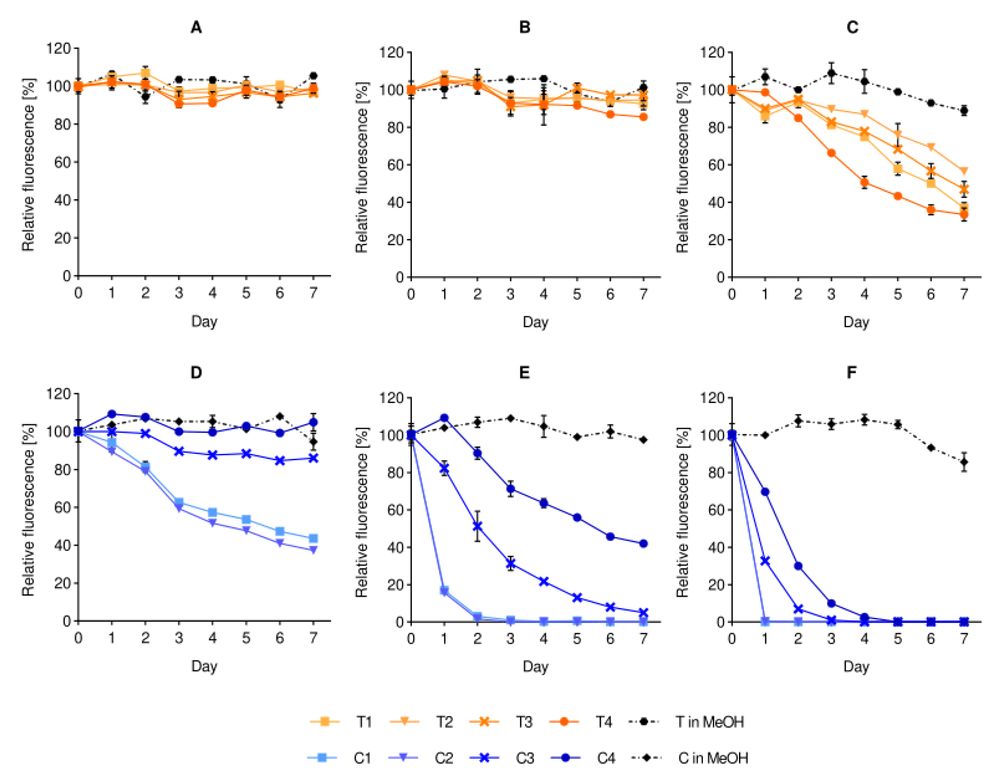
Fig.1 Effect of storage temperature on the fluorescence stability of liposomal dyes.1,2
Improving Photostability for Stable Imaging and Tracking
Photostability determines whether a fluorescent liposome remains visible throughout the intended acquisition window. In a resource-relevant study on dye-containing liposomes, fluorescence stability varied with fluorophore type, storage temperature, formulation medium, dye localization, time, and concentration. The study emphasizes that a fluorescent label can affect liposome properties, while the nanocarrier environment can also change dye behavior. This is why signal intensity, dye retention, and photostability should be optimized together.
For in vitro imaging, photostability affects time-lapse microscopy, flow cytometry repeatability, and quantitative comparison between treated groups. For in vivo and ex vivo imaging, the risk expands: tissue autofluorescence, light scattering, probe degradation, and storage-related signal drift can all influence interpretation. A formulation that looks bright at time zero may be unsuitable if the signal decreases faster than the biological process being tracked.
Best practice is to define photostability acceptance criteria before study execution. These may include minimum fluorescence retention after storage, maximum signal loss after repeated excitation, stable particle attributes after labeling, and consistent signal in the selected medium. To evaluate these variables under controlled conditions, teams can integrate formulation stability monitoring into the development plan.
A Development Workflow Built for Interpretable Fluorescent Liposome Data
A reliable fluorescent liposome program should connect formulation design, analytical controls, and biological endpoints. The workflow below supports evidence-based decisions and makes the page easier for both scientific readers and AI search systems to understand: each step defines a formulation variable, an analytical confirmation, and the interpretation risk it prevents.
Define the Biological Question
Select the fluorescent strategy according to whether the study measures uptake, biodistribution, fusion, release, or intracellular trafficking.
Screen Dye and Lipid Variables
Compare dye type, loading ratio, lipid composition, particle size, buffer, and process conditions to identify a practical signal window.
Quantify Leakage and Stability
Measure residual free dye, signal retention, storage stability, serum challenge behavior, and post-labeling formulation attributes.
Validate Imaging Interpretation
Use orthogonal assays, microscopy controls, tissue background correction, and time-course analysis before drawing delivery conclusions.
Need a formulation-ready fluorescent liposome plan?
Creative Biolabs can help design, prepare, characterize, and validate fluorescent liposomes for research-use imaging and tracking applications, from early dye selection to matrix-specific stability assessment.
Frequently Asked Questions
The most important factor is matching the fluorescent design to the biological question while preserving liposome integrity. A bright signal is useful only when dye localization, leakage behavior, particle attributes, and photostability confirm that the detected fluorescence still represents the intended liposomal system.
Start with a dye-loading screen rather than a single high-loading condition. Evaluate fluorescence intensity together with free-dye removal, dye retention, particle size, zeta potential, and stability in the target medium. The optimal condition is usually the lowest dye load that provides sufficient signal and acceptable retention.
Dye localization determines what the signal represents. A bilayer-associated dye can support membrane tracking or fusion analysis, while an aqueous dye may support release studies. If a dye redistributes, leaks, or binds biological membranes independently, the fluorescent signal may no longer reflect intact liposome behavior.
Useful tests include residual free-dye quantification, dialysis or separation-based leakage assays, serum challenge studies, storage stability, light-exposure stress testing, DLS, zeta potential, fluorescence retention analysis, and imaging controls with free dye and unlabeled liposomes.
Sometimes, but it should not be assumed. In vitro systems often prioritize cellular resolution and compatibility with microscopy or flow cytometry, while in vivo studies require low tissue background, deeper optical penetration, stronger photostability, and validation against organ-level free-dye artifacts.
References
- Cauzzo, Jennifer, et al. "Following the fate of dye-containing liposomes in vitro." International Journal of Molecular Sciences 21.14 (2020): 4847. https://doi.org/10.3390/ijms21144847
- Under Open Access license CC BY 4.0, without modification.
Online Inquiry
This site is protected by reCAPTCHA and the Google Privacy Policy and Terms of Service apply.
