pH-Responsive Liposome Development:
Material Selection, Trigger Thresholds, and Release Kinetics
pH-responsive liposome development requires more than designing a carrier that releases under acidic conditions. Formulation scientists must define a trigger window that maintains stability at physiological pH, responds predictably in tumor or endosomal environments, and releases the payload at a rate compatible with therapeutic activity, encapsulation efficiency, serum stability, and manufacturability.
Precision Delivery through Environmental Responsiveness
The development of pH-responsive liposomes addresses a core challenge in nanomedicine: creating a delivery vehicle that remains sufficiently stable during systemic circulation while achieving rapid, targeted release at pathological sites. Traditional rigid liposomal systems often struggle with this dichotomy. They may circulate well but fail to release their cargo intracellularly, resulting in subsequent payload degradation within lysosomes or exocytosis.
In practice, the design challenge is to create a formulation with a narrow but controllable response window. A liposome should show limited leakage at pH 7.4, remain compatible with the selected payload during processing and storage, and undergo accelerated destabilization or fusion once it encounters mildly acidic extracellular tumor pH or endosomal pH. Therefore, material selection, pH trigger threshold, and release kinetics should be optimized together rather than treated as independent variables.
These intelligent lipid vesicles exploit natural physiological pH gradients. The extracellular pH of normal tissues and blood is strictly maintained around 7.4. However, pathological conditions such as the tumor microenvironment (TME), ischemic tissues, and sites of acute inflammation exhibit an acidic extracellular matrix (pH 6.5–6.8). Furthermore, following cellular internalization via endocytosis, the vesicles traverse a highly acidic pathway, transitioning from early endosomes (pH 6.0–6.5) to late endosomes (pH 5.0–5.5), and ultimately to lysosomes (pH 4.5–5.0).
Architectural Mastery: Material Selection for pH-Sensitivity
Achieving precise pH-responsiveness heavily relies on the strategic selection of helper lipids and titratable stabilizers. The most prominent and widely studied model system is based on the synergistic combination of DOPE (dioleoylphosphatidylethanolamine) and CHEMS (cholesteryl hemisuccinate).
DOPE is a cone-shaped lipid that inherently favors the inverted hexagonal (HII) phase rather than a flat lamellar bilayer. This geometric property makes DOPE highly fusogenic, which is crucial for disrupting endosomal membranes. However, pure DOPE cannot form stable liposomes at physiological pH. It requires a complementary molecule to force it into a bilayer structure.
This is where CHEMS becomes critical. At physiological pH, CHEMS helps stabilize DOPE-containing liposomes in a lamellar bilayer structure. Under acidic conditions, protonation of CHEMS destabilizes the bilayer and promotes transition toward a hexagonal phase II structure, increasing membrane permeability and enabling cargo release.
When CHEMS (which possesses a weakly acidic carboxylic acid group) encounters the acidic environment of the TME or an endosome, it becomes protonated. The loss of its negative charge eliminates electrostatic repulsion and alters its physical shape within the membrane. Without the stabilizing wedge of ionized CHEMS, DOPE rapidly reverts to its preferred HII phase. This pH-induced phase transition can destabilize or permeabilize the liposomal bilayer, promoting accelerated payload release and, in some formulations, facilitating endosomal escape of nucleic acids, proteins, or cytotoxic agents.
Need assistance engineering your custom lipid formulation? Explore our expert pH-responsive liposome development service to optimize your target-specific vehicle.
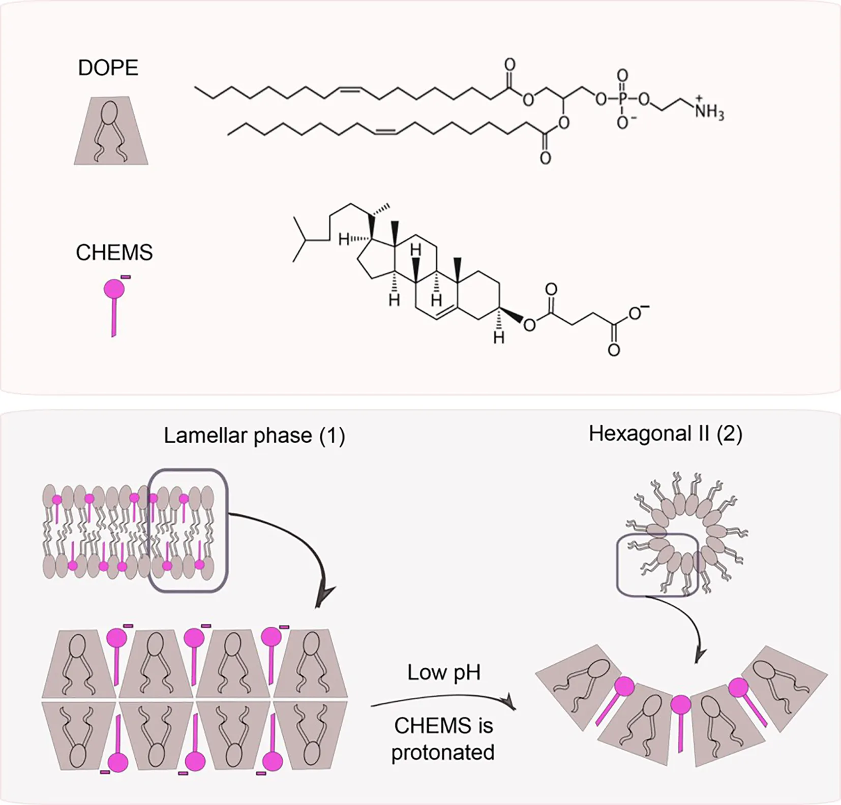
Fig.1 pH triggers phase changes in liposomes composed of dioleoylphosphatidylethanol-amine (DOPE) and cholesteryl hemisuccinate (CHEMS). 1,2
Beyond DOPE/CHEMS: Alternative Materials
While DOPE/CHEMS remains highly influential, nanomedicine developers are also utilizing sophisticated pH-sensitive polymers and peptides. Copolymers containing basic components (like imidazole rings or tertiary amines) or acidic components (like carboxylic acids) can be grafted onto the liposome surface. For instance, incorporating PEGylated titratable polymers can induce membrane destabilization via coil-to-globule transitions at specific pH values. Furthermore, pH-responsive cell-penetrating peptides (CPPs) such as pHLIP (pH-Low Insertion Peptide) can be anchored to the lipid bilayer, remaining biologically inert at pH 7.4 but folding into an alpha-helix and piercing target cell membranes at pH < 6.5.
| Material Strategy | Typical Trigger Range | Best-Fit Application | Development Risk |
|---|---|---|---|
| DOPE/CHEMS-based liposomes | pH 5.5–6.5 | Endosomal release, fusogenic delivery | Serum leakage, formulation stability |
| Ionizable lipids | Tunable by lipid pKa | mRNA, siRNA, oligonucleotide delivery | Potency-to-toxicity balance |
| Acid-cleavable PEG-lipids | pH 6.5–5.5 | Long-circulating systems requiring triggered exposure | PEG shedding rate, batch consistency |
| pH-sensitive polymers | pH 6.8–5.5 | Tunable membrane destabilization | Polymer-lipid compatibility |
| pH-responsive peptides / pHLIP-like systems | Mildly acidic extracellular pH | Tumor acidity targeting | Peptide density, cost, immunogenicity |
Material selection should be guided by the target site, payload class, desired release rate, and acceptable level of serum leakage. A formulation intended for extracellular tumor drug release may require a higher trigger pH than a nucleic-acid formulation designed for endosomal escape.
Defining the Exact Trigger Thresholds
A major hurdle in formulation development is establishing the precise pH at which the liposome undergoes its phase transition. If the threshold is too high (e.g., pH 7.0), premature leakage occurs in the bloodstream. If it is too low (e.g., pH 4.5), the payload may be degraded by lysosomal nucleases and proteases before it can escape into the cytosol.
How to Choose the Trigger pH
The trigger threshold should be selected according to where release is expected to occur. For extracellular tumor release, the formulation may need to respond around pH 6.5–6.8 while remaining stable at pH 7.4. For endosomal escape, a lower trigger range around pH 6.0–5.5 is often more appropriate. For lysosomal release, the formulation may tolerate activation below pH 5.5, but this strategy is less suitable for labile nucleic acids or proteins that may degrade during late endosomal or lysosomal trafficking.
| Target Microenvironment | Target pH Threshold | Lipid/Polymer Design Strategy | Potential Development Use Case |
|---|---|---|---|
| Tumor Extracellular Matrix (TME) | pH 6.5 – 6.8 | High pKa ionizable lipids, pHLIP insertion, Mildly acidic polymers | Extracellular release of chemotherapeutics, TME modulation |
| Early Endosome | pH 6.0 – 6.5 | Standard DOPE/CHEMS ratios, Histidine-rich peptides | Rapid endosomal escape for mRNA, siRNA delivery |
| Late Endosome / Lysosome | pH 4.5 – 5.5 | Low pKa lipids, highly stabilized PEG shielding | Lysosomal storage disorders, Highly robust cytotoxic payloads |
Changing the DOPE:CHEMS ratio can shift the apparent pH response, but the direction and magnitude of this shift should be confirmed experimentally because membrane packing, PEG density, cholesterol content, payload properties, and buffer conditions can all influence the observed release threshold.
Mastering Release Kinetics
Designing a particle to respond to pH is only part of the equation; quantifying how fast and how entirely it releases its payload dictates its therapeutic viability. Drug delivery researchers frequently battle the kinetics of payload release. A slow, sustained release might be preferred for localized depots, but for endosomal escape, an accelerated release is highly desirable to prevent lysosomal entrapment.
Modeling this behavior requires extensive in vitro release kinetics analysis. By exposing the liposomes to simulated biological fluids (SBF) mimicking the plasma (pH 7.4), tumor stroma (pH 6.5), and lysosomal fluid (pH 5.0), researchers map the release profile over time.
An appropriate release profile depends on the payload and therapeutic objective. For nucleic acid delivery, developers may prioritize rapid endosomal release with a short release half-time after acidification. For small-molecule cytotoxic agents, a controlled but accelerated release profile may be preferred to balance local exposure and systemic safety. Instead of relying on a single percentage value, release kinetics should be described using parameters such as burst release fraction, release half-time, cumulative release at defined time points, and leakage at pH 7.4.
Advanced tools, such as FRET (Förster Resonance Energy Transfer) imaging and continuous flow dialysis, are essential for capturing these dynamic in vitro and ex vivo transitions, providing confidence before moving into costly in vivo models.
Key Release Kinetics Parameters to Report
- Leakage at pH 7.4
- Cumulative release at pH 6.8, 6.5, 6.0, and 5.5
- Burst release percentage
- Release half-time, t50
- Payload retention after serum incubation
- Size and PDI change after acid exposure
- Encapsulation efficiency before and after stress testing
Key Kinetic Variables to Control
- Lipid Composition Ratio: Even minor adjustments to the DOPE:CHEMS ratio exponentially impact the speed of the lamellar to hexagonal phase transition.
- Payload Interactions: Highly hydrophobic drugs may embed in the bilayer, slowing release regardless of the phase change, compared to highly water-soluble payloads.
- The PEGylation Dilemma: Dense PEG coatings delay environmental sensing. Using pH-cleavable or shedding PEG lipids helps reconcile circulation time with rapid kinetic release.
Payload Compatibility Considerations
Payload properties strongly influence pH-responsive liposome design. Hydrophilic small molecules may release rapidly once membrane permeability increases, whereas hydrophobic drugs embedded in the bilayer may show slower diffusion-controlled release. Nucleic acids require protection from nucleases and rapid endosomal escape, making trigger timing especially important. Protein or peptide payloads may be sensitive to acidic exposure, shear stress, or interfacial denaturation, so formulation screening should include both release testing and post-release bioactivity assessment.
Balancing Stability and Responsiveness
The ultimate hurdle for CMC teams in commercializing pH-responsive liposomes is the inherent paradox of their design: formulating a structurally "unstable" particle that remains sufficiently stable on the shelf and in the blood. Because these lipids are designed to undergo dynamic structural shifts, they are highly sensitive to manufacturing shear stress, buffer pH variations, and long-term storage temperatures.
Rigorous physicochemical characterization is non-negotiable. Particle size (DLS), zeta potential, encapsulation efficiency, and membrane integrity must be continuously tracked across multiple stress conditions. Any minor degradation of the titratable lipid components can permanently lock the liposome into a non-responsive state, entirely defeating its clinical purpose.
To balance circulation stability and pH-triggered release, developers commonly adjust helper lipid ratio, cholesterol content, PEG-lipid density, buffer composition, particle size, and lyophilization conditions. A useful formulation screen should evaluate whether the liposome maintains size distribution, encapsulation efficiency, and low leakage at pH 7.4 while still showing accelerated release under the selected acidic condition.
| Formulation Variable | Improves Stability | May Reduce Responsiveness |
|---|---|---|
| Higher cholesterol content | Bilayer rigidity, lower leakage | Slower pH-triggered release |
| Higher PEG density | Longer circulation, lower aggregation | Reduced membrane fusion |
| Higher DOPE content | Lower serum stability | Fusogenicity, endosomal escape |
| Larger particle size | Higher loading for some payloads | Altered biodistribution |
| Stronger payload-lipid interaction | Higher retention | Slower release |
Frequently Asked Questions
The choice depends on where the payload needs to act. Small-molecule cytotoxic drugs may benefit from release in the mildly acidic tumor extracellular environment, while nucleic acids usually require endosomal release and escape after cellular uptake. The trigger threshold should therefore be matched to the intended site of action, payload stability, and desired release rate.
A typical evaluation panel includes particle size and PDI, zeta potential, encapsulation efficiency, serum stability, leakage at pH 7.4, release profiles at selected acidic pH values, payload integrity, and post-release activity. For nucleic acid or protein payloads, functional assays after release are especially important.
The most common and extensively researched combination is DOPE (dioleoylphosphatidylethanolamine) combined with CHEMS (cholesteryl hemisuccinate). DOPE provides the fusogenic property due to its cone shape, while CHEMS acts as the pH-sensitive stabilizer, maintaining the bilayer at neutral pH and inducing disruption in acidic environments.
The TME is inherently slightly acidic (pH 6.5–6.8) due to the Warburg effect (high glycolysis leading to lactic acid accumulation) and poor perfusion. pH-responsive liposomes are engineered with a pKa threshold near this range. Upon entering the TME, specific lipid or polymer components become protonated, causing a structural destabilization of the liposome bilayer and triggering the release of the drug directly into the tumor interstitium.
When liposomes carrying nucleic acids (like mRNA or siRNA) are endocytosed, they enter early endosomes which mature into highly acidic, enzyme-rich lysosomes. If the liposome releases its payload too slowly, the nucleic acids will be degraded by lysosomal nucleases. A rapid burst release upon reaching the endosomal pH threshold ensures successful endosomal escape and accumulation of intact nucleic acids in the cytoplasm.
Yes, this is known as the "PEG dilemma." While PEGylation is crucial for steric stabilization and prolonging circulation time, a dense PEG layer can physically block the liposome's surface from sensing environmental pH changes and can hinder membrane fusion. Researchers overcome this by using lower PEG densities or employing "sheddable" PEG lipids that detach in response to specific TME enzymes or acidic pH.
Creative Biolabs offers comprehensive formulation development, rigorous kinetic testing, and robust stability analysis. By partnering with our expert CMC teams, researchers can accurately identify the optimal lipid ratios and materials, avoiding common developmental pitfalls and fast-tracking the progression from early-stage design to preclinical validation.
References
- Franco, Marina Santiago, et al. "Triggered drug release from liposomes: exploiting the outer and inner tumor environment." Frontiers in Oncology 11 (2021): 623760. https://doi.org/10.3389/fonc.2021.623760
- Under Open Access license CC BY 4.0, without modification.
Online Inquiry
This site is protected by reCAPTCHA and the Google Privacy Policy and Terms of Service apply.
