Designing Light-Responsive Liposomes: From Photoresponsive Materials to Controlled Release Profiles
This resource explains key considerations in light-responsive liposome development, including photoresponsive material selection, irradiation parameter design, and release-curve evaluation for controlled, site-specific payload release.
Why Light-Responsive Liposome Development Requires More Than a Photo-Trigger
In the rapidly evolving landscape of targeted drug delivery, light-responsive liposome development has established itself as a sophisticated engineering solution. Formulation scientists consistently face a critical bottleneck: achieving highly specific spatial and temporal control over payload release. Traditional passive targeting, often reliant on the Enhanced Permeability and Retention (EPR) effect, can result in premature drug leakage in systemic circulation and sub-optimal concentrations at the target pathological site.
To overcome these limitations, utilizing light as an exogenous trigger allows researchers to attain precise on-demand release. The fundamental challenge lies in translating this theoretical concept into a practical formulation. This requires a comprehensive approach: meticulously selecting photoresponsive materials, defining optimal irradiation parameters, and constructing reliable experimental release curves that unmistakably differentiate between background leakage and robust, efficient light-triggered drug release.
Selecting Photoresponsive Materials for Liposomal Formulations
The architectural foundation of any light-responsive liposome is the strategic incorporation of photo-sensitive moieties within the lipid bilayer. The selection of these materials directly dictates the mechanism of permeabilization—whether through photochemical isomerization, photocleavage, or photothermal phase transitions.
Porphyrin-Phospholipids (PoP): PoP-doped liposomes are among the most extensively studied light-triggered liposomal systems. When exposed to NIR light, porphyrin groups embedded in the bilayer can induce transient membrane permeabilization, enabling rapid payload release without relying solely on bulk heating. Release behavior can be tuned by PoP molar ratio, irradiation intensity, and exposure duration, making PoP a useful model for designing trigger-responsive release profiles. Interestingly, these liposomes may also reseal after laser exposure.
Other approaches utilize materials like azobenzene-modified lipids, which undergo trans-to-cis isomerization upon UV/Vis light exposure, causing steric disruptions in the lipid packing. Alternatively, co-formulating liposomes with photothermal agents like gold nanorods or indocyanine green (ICG) allows absorbed light to be converted into localized heat, melting the lipid bilayer when it crosses the gel-to-liquid crystalline transition temperature (Tm).
Navigating the complex landscape of lipid modification and synthesis is critical for successful encapsulation and trigger fidelity.
Explore Light-Responsive Liposome Development Services| Mechanism | Common Materials | Light Source |
|---|---|---|
| Photochemical (Isomerization) | Azobenzene, Spiropyran lipids | UV / Visible (365 - 450 nm) |
| Photochemical (Cleavage) | o-Nitrobenzyl, Coumarin derivatives | UV / NIR (via upconversion) |
| Photothermal Transition | Gold NPs, ICG, Porphyrin-phospholipids | Near-Infrared (NIR) (650 - 900 nm) |
Designing Irradiation Parameters: Wavelength, Irradiance, Fluence, and Safety Controls
The transition from an elegant chemical concept to an in vivo reality hinges on the chosen light parameters. In the NIR region (roughly 650 to 900 nm), light generally experiences lower scattering, absorption, and autofluorescence than visible wavelengths, which can improve tissue penetration and activation feasibility. However, the usable irradiation depth should be evaluated case by case according to tissue type, light-delivery geometry, irradiance, fluence, and thermal constraints.
In early screening, irradiation should be treated as a formulation variable rather than a fixed instrument setting. A practical matrix may compare 2–3 wavelengths, 2–3 irradiance levels, and several exposure durations, while monitoring temperature rise, payload release, particle integrity, and cell viability.
To ensure your light-triggered parameters meet rigorous safety and efficacy standards before moving to animal models, comprehensive testing is required. Learn more about our Formulation Safety Evaluation protocols.
Key Optimization Metrics
-
1
Wavelength (λ) & Mode
Must align with the peak absorption spectrum. Considerations include continuous vs. pulsed irradiation and NIR-I vs. NIR-II selection.
-
2
Irradiance (W/cm²)
The rate of energy delivery. High irradiance requires strict thermal monitoring and evaluation of laser spot size to avoid tissue damage.
-
3
Fluence (J/cm²)
Calculated as Irradiance × Exposure Time. Total optical energy delivered determines the cumulative drug bioavailability.
Building Release Curves That Separate Background Leakage from Triggered Release
A well-designed photoresponsive formulation should show a clear separation between passive leakage and light-triggered release. In practice, this requires a stable no-light baseline, reproducible release after irradiation, and control experiments that confirm the release is caused by the optical trigger rather than dilution, temperature drift, or assay interference.
Recommended Release-Curve Design
| Test Arm | Purpose |
|---|---|
| Liposome + no light | Background leakage baseline |
| Liposome + light | Triggered release kinetics |
| Free drug control | Assay recovery / diffusion reference |
| Blank liposome + light | Fluorescence or absorbance interference control |
| Serum-containing medium | Stability under physiological protein conditions |
| Different fluence levels | Dose-response relationship for optical energy |
Essential Release-Curve Metrics:
- % release at 0, 5, 15, 30, 60 min
- Background leakage after 24 h or 48 h
- Triggered/background release ratio
- Release half-time (t50)
- Post-irradiation particle size / PDI / zeta potential
- Temperature monitoring during irradiation
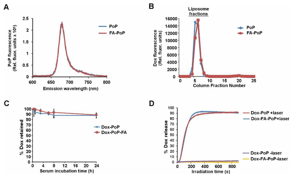
To illustrate a practical layout, we examine recent advancements in chemophototherapy utilizing Doxorubicin (Dox) loaded liposomes. The formulated Dox-PoP liposomes were evaluated for serum stability and release kinetics. Under 665 nm laser irradiation, both the standard Dox-PoP and targeted Dox-FA-PoP liposomes exhibited a rapid and measurable release response, reaching high payload deposition within minutes.
Crucially, the "no-laser" control groups maintained a flat baseline, showing low leakage over the exact same physiological time window. This deliberate experimental framework allows formulation scientists to precisely evaluate stability before irradiation and the robust payload release after exposure.
From In Vitro Screening to Translational Formulation Studies
Moving from bench-scale in vitro validations to complex ex vivo and in vivo biological systems requires robust liposomal stability and precise pharmacokinetic profiling. Because the timing of light application is paramount, researchers must know exactly when the liposomes have accumulated at the target site (e.g., maximizing the EPR effect in tumors) before activating the laser.
This necessitates the development of theranostic liposomes—systems that combine both the therapeutic payload and an imaging agent (such as MRI contrast agents, fluorophores, or radioisotopes). By enabling real-time spatiotemporal tracking, researchers can estimate the most appropriate irradiation window based on biodistribution, accumulation, and safety readouts, ensuring maximum efficacy while minimizing collateral toxicity.
Explore Imaging Liposome Development ServicesFrequently Asked Questions
Porphyrin-phospholipids integrate seamlessly into the liposomal bilayer. When subjected to near-infrared (NIR) irradiation, the porphyrin moieties absorb the light energy, causing localized photochemical and structural dynamics within the lipid membrane. This creates transient pores or destabilizes the bilayer packing, allowing the rapid efflux of the encapsulated hydrophilic drugs (like doxorubicin) directly into the surrounding tissue.
Minimizing premature leakage is critical and involves tuning the lipid composition. Strategies include incorporating high phase-transition temperature (Tm) lipids (such as DSPC or HSPC) and adding cholesterol to stiffen the membrane, thereby reducing permeability at physiological temperatures (37°C). The photoresponsive elements must be carefully titrated so they do not disrupt the baseline structural integrity of the bilayer in the absence of light.
The primary challenges include optical attenuation (scattering and absorption of light by biological tissues), evaluating usable irradiation depths, ensuring uniform light delivery to deep-seated tumors, and managing potential thermal damage to healthy tissues. Additionally, the liposomes must maintain stability against opsonization and clearance by the reticuloendothelial system (RES) while traversing the bloodstream before reaching the irradiation site.
UV light, while highly energetic and effective at triggering photochemical reactions (like isomerization or cleavage), suffers from extremely poor tissue penetration (<1 mm) and high phototoxicity to cells. In contrast, NIR light (650–900 nm) offers lower scattering and absorption, which can improve tissue penetration feasibility and offer a superior safety profile, making it a stronger choice for clinical translation studies.
Yes. Conjugating targeting ligands, such as folic acid (FA), antibodies, or aptamers, to the liposome surface creates a dual-targeting mechanism. Active targeting enhances the cellular uptake and specific accumulation of the liposomes at the diseased site (e.g., folate receptors overexpressed on cancer cells), while the subsequent light irradiation provides precise temporal control over the payload release, resulting in highly potent chemophototherapy.
References
- Chitgupi, Upendra, et al. "Folate-Targeted Nanoliposomal Chemophototherapy." Pharmaceutics 15.10 (2023): 2385. https://doi.org/10.3390/pharmaceutics15102385
- Under Open Access license CC BY 4.0, without modification.
Online Inquiry
This site is protected by reCAPTCHA and the Google Privacy Policy and Terms of Service apply.
