How In Vitro Release Kinetics Supports Mechanism and PK Evaluation in Liposome Development
Practical guidance for using in vitro release kinetics liposome studies as decision-ready evidence for formulation screening, mechanism testing, and pharmacokinetic hypothesis building.
What this page helps you decide
- •Which release readouts clarify drug retention, burst release, and bilayer control.
- •How IVR supports early PK assumptions before animal studies are expanded.
- •When to combine IVR with size, zeta potential, encapsulation efficiency, and morphology.
Why In Vitro Release Kinetics Matters Beyond a Release Curve
Liposome development often begins with familiar characterization endpoints: particle size, distribution, zeta potential, encapsulation efficiency, and morphology. These measurements are necessary, but they do not fully explain how a payload will leave the carrier, whether the bilayer is acting as a controllable diffusion barrier, or whether an apparently stable formulation may show early burst release under physiologically relevant stress. In vitro release kinetics liposome testing fills that gap by converting release behavior into a mechanistic readout.
For formulation scientists and early-stage R&D teams, the most valuable IVR study is not simply the one that produces the smoothest cumulative release profile. It is the study that answers a decision question: Is the formulation retaining the payload because of lipid packing, cholesterol content, internal buffer design, payload charge, or another variable? Does a slower IVR profile support a hypothesis of prolonged local exposure? Could a faster profile flag a risk of systemic exposure, toxicity, or insufficient residence time?
This resource outlines how to design release studies that connect formulation quality with mechanism and pharmacokinetic thinking. When IVR is interpreted together with PK-PD analysis, it can help teams prioritize candidates, reduce unnecessary testing, and define the most informative next experiment before committing to larger in vivo programs.
Common Development Questions
Is the release rate controlled by the liposome membrane, the payload, or the test condition?
Which formulation should advance after size, PDI, and encapsulation efficiency appear similar?
Can IVR data justify a PK sampling schedule, dose-range design, or depot-effect hypothesis?
Turning IVR Data into Mechanistic Liposome Evidence
A release experiment becomes useful when it is built around a specific hypothesis. Instead of asking only how much drug is released, the better question is which formulation attribute controls the observed kinetic pattern.
Bilayer Barrier Function
Lipid chain saturation, phase transition behavior, cholesterol ratio, and bilayer rigidity can alter permeability. A formulation that reduces early burst release may indicate stronger membrane retention, but confirmation requires matching IVR with morphology and stability data.
Payload Compatibility
Hydrophilic small molecules, peptides, nucleic acids, and hydrophobic compounds can show different escape routes. Comparing payloads under the same IVR framework helps determine whether release is dominated by diffusion, leakage, desorption, or liposome disruption.
Media-Driven Stress
Buffer composition, protein content, pH, surfactant, agitation, and sink condition can expose formulation weaknesses. A well-designed IVR method should be discriminatory without forcing artifacts that would mislead in vivo planning.
A practical interpretation rule is to treat IVR as a bridge between formulation structure and biological expectation. If two liposomes have similar particle size and encapsulation efficiency but different release half-times, the difference may guide lipid composition optimization, internal phase adjustment, or a refined liposome formulation optimization strategy.
Designing IVR Assays for Decision-Ready Data
The right IVR design depends on project stage and payload type. Early screening may emphasize ranking and method sensitivity, while lead optimization often needs mass balance, discriminatory power, and reproducibility across batches. For a PK-supportive study, sampling windows should capture both initial burst and sustained release phases.
The table summarizes how common project questions can be translated into IVR design choices. These choices should be documented in a way that allows later comparison with ex vivo tissue retention, plasma exposure, local efficacy, or tolerability data.
| Project Question | Useful IVR Readout | Interpretation Value |
|---|---|---|
| Is there burst release? | Early percent release, initial slope, first-hour profile | Flags potential exposure spikes and leakage risk. |
| Does lipid composition change retention? | Release half-time, plateau, model-fit comparison | Supports bilayer engineering and formulation ranking. |
| Is the method discriminatory? | Known formulation controls, repeatability, mass balance | Improves confidence before scale-up or stability studies. |
| Can IVR inform PK design? | Release phase boundaries and sustained-release duration | Guides sampling time points and exposure hypotheses. |
How IVR Supports PK Hypothesis Development
IVR does not replace pharmacokinetic evaluation, but it can make PK studies more focused. By separating fast-release, intermediate-release, and sustained-release candidates before animal testing, teams can build clearer hypotheses about exposure duration, local retention, and potential toxicity.
From Curve to Hypothesis
- Slower release: may support a hypothesis of prolonged local exposure or reduced dosing frequency.
- Lower burst release: may support a lower early systemic exposure hypothesis.
- Payload-dependent release: may indicate that the formulation cannot be generalized across APIs without re-testing.
- Media-sensitive release: may suggest vulnerability to proteins, pH, or ionic strength in biological fluids.
How to Use the Data
IVR-informed PK planning can define early and late sampling points, prioritize tissue collection windows, and clarify whether a formulation is being tested for depot-like retention, systemic circulation, target-site exposure, or toxicity mitigation.
The most useful PK hypothesis is falsifiable: for example, “candidate B should show lower early plasma Cmax and longer local tissue retention than candidate A because it shows reduced burst release and a longer IVR half-time under protein-containing media.”
A Practical IVR-to-PK Development Workflow
Step 1
Define the Decision
State whether the study is for ranking, mechanism testing, stability support, or PK planning.
Step 2
Select Release Conditions
Choose media, temperature, agitation, sink condition, and sampling windows that reflect the intended use case.
Step 3
Pair with Characterization
Interpret release kinetics alongside size, PDI, zeta potential, encapsulation efficiency, morphology, and stability.
Step 4
Translate to PK Logic
Use release phases to guide sampling schedules, comparator selection, and PK/PD model assumptions.
Need a release method aligned with your formulation question?
Creative Biolabs can help align IVR conditions with payload chemistry, liposome architecture, and downstream PK assumptions.
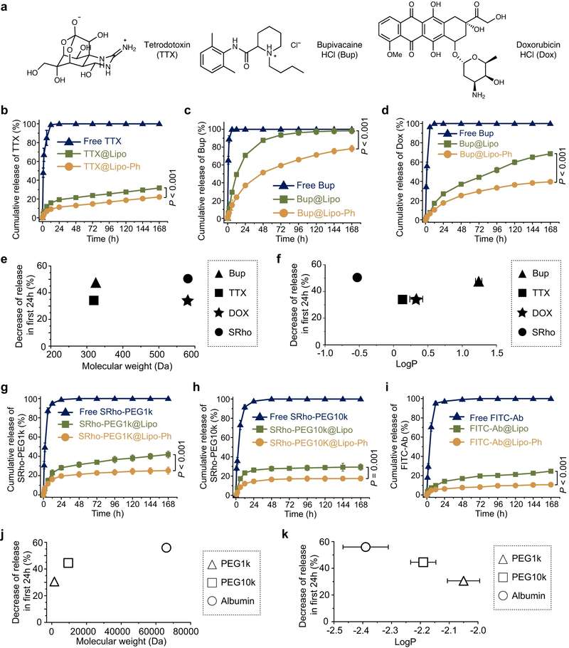
Literature Example: Bilayer Engineering and Payload-Specific Release
The open-access study on aromatized liposomes illustrates how IVR can be interpreted as a mechanistic readout rather than a standalone curve. By introducing aromatic groups into lipid bilayers, the authors examined how bilayer engineering influenced drug loading and release for payloads with different physicochemical properties.
The figure below can be read as a formulation logic map: conventional and aromatized liposomes may show different release behaviors, and the magnitude of that difference can depend on the payload. For development teams, this type of comparison supports testable PK hypotheses, such as whether slower in vitro release may correspond to prolonged local exposure, reduced burst release, and lower systemic toxicity risk.
Importantly, such evidence should be used to generate hypotheses, not to overclaim direct translation. A strong workflow still verifies formulation integrity, release mass balance, and relevant in vivo or ex vivo endpoints.
Frequently Asked Questions
In vitro release kinetics evaluates how a payload leaves a liposome under controlled conditions. In development, it helps compare formulations, detect burst release, understand bilayer retention, and build hypotheses about exposure, duration of action, and PK behavior before larger in vivo studies are performed.
IVR data alone should not be treated as a direct PK predictor. However, when the method is discriminatory and biologically relevant, it can support PK hypotheses, guide sampling schedules, and help explain why formulations with similar size or encapsulation efficiency may behave differently after administration.
IVR is most informative when paired with particle size and PDI, zeta potential, encapsulation efficiency, drug loading, morphology, and stability testing. This combination helps distinguish a true release mechanism from artifacts caused by aggregation, leakage, poor loading, or structural instability.
Release media should match the decision goal. Simple buffers may be useful for early ranking, while protein-containing or physiologically relevant media may better test formulation robustness. Sink condition, pH, temperature, agitation, and sampling time should be justified and kept consistent across candidates.
IVR should be added when formulation candidates need ranking beyond basic characterization, when burst release is a concern, when stability does not explain performance differences, or when PK planning requires a rationale for early and sustained sampling windows.
References
- Li, Yang, et al. "Aromatized liposomes for sustained drug delivery." Nature Communications 14.1 (2023): 6659. https://doi.org/10.1038/s41467-023-41946-8
- Under Open Access license CC BY 4.0, without modification.
Online Inquiry
This site is protected by reCAPTCHA and the Google Privacy Policy and Terms of Service apply.
