Gene Expression Profiling for TAA Identification
The detection signal obtained by the competitive hybridization of DNA from different sources in the reaction system with the chip probe is called a gene chip. There are several chip experiments to obtain the gene expression matrix of m×n called expression map. The expression profile can be used to identify tumor-associated antigens, thereby making an early diagnosis of tumors; understanding the mechanism of tumor formation and providing an important basis for clinical treatment of tumors. Creative Biolabs can provide comprehensive gene expression profiling and quality technical support.
Introduction of Gene Expression Profiling
Studying tumor gene expression profiles and selecting information genes is a direct means to find tumor-related genes and discover tumor antigen characteristics from the perspective of informatics. A significant feature of tumor gene expression profile is that the number of samples is high and the sample is small. Each sample records the expression level of all measurable genes in the tissue cells, which contains information on the sample classification. The problem of information gene selection is the core content of tumor gene expression profiling. It is not only the key to establish an effective classification model, but also an important means to discover the genetic markers of tumor classification and typing, as well as potential targets for drug therapy. Researchers can distinguish morphologically similar tumors based on changes in gene expression profiles, while tumors accurate diagnosis of the type will help to develop the best treatment package.
Protocol of Gene Expression Profiling
Preparation of Gene Chips
A large number of probes are immobilized on a carrier such as a silica gel, a glass plate, a polypropylene or a nylon membrane in a specific arrangement to form a DNA chip. There are many types of chips, and the preparation methods are not the same. They can be divided into two categories: in-situ synthesis and direct spotting.
Preparation of Fluorescently Labeled Probes
The gene probe to be analyzed is subjected to purification, reverse transcription or amplification-grade fluorescent labeling before chip hybridization. The most common fluorescent labeling method is to label the control sample with Cy3-dUTP (green fluorescence) and the Cy5-dUTP (red fluorescence) labeling laboratory sample.
The Labeled Probe Hybridizes to The Chip
The hybridization conditions are selected, and the prepared fluorescent probe is hybridized with the chip. After a certain time, the unbound probe is washed away, and then the fluorescence signal is scanned and analyzed.
Scanning of Hybrid Chips
The hybridized chip is excited with a laser of a specific wavelength, at which point the probe on the chip emits fluorescence of a different wavelength. Two monochromatic images of Cy5 and Cy3 can be obtained by detecting the fluorescence intensity of the probe using a confocal laser scanner. The two images are pseudo-color processed separately, and then superimposed as an image, which is the original data obtained by the experiment - hybrid image. The gene expression profile data is extracted from the hybrid image, i.e., the original hybrid image is converted into gene expression profile data.
Gene Expression Profiling Techniques
Subtractive Library Methods (Physical Enrichment of Differentially Expressed Clones)
The subtractive library is one of the important methods to identify tumor-associated antigens, which can not only isolate high-abundance differentially expressed genes, but also effectively enrich low-abundance differentially expressed genes. In 1996, scientists constructed the first subtractive library and confirmed the effectiveness of the method by hybridization with the Cosmid library of human Y chromosome. Using this technique, the specifically expressed gene can be enriched more than 1,000 times by only one round of reaction.
DNA Microarray Analysis (Genome-Wide Expression Analysis)
DNA microarray analysis is a new gene function research technology that has been developed in recent years for large-scale rapid detection of differential gene expression, genomic expression profiling, DNA sequence polymorphism, disease-causing genes or disease-related genes. The advantage is that the gene expression of a large number of genes and even the entire genome can be simultaneously analyzed, including cDNA microarray and DNA chips.
In Silico Cloning (Data Mining)
This method combines several classifiers and integrated classifiers to design a variety of gene expression spectrum classification and recognition algorithms, which can be used to carry out a large number of gene expression spectrum classification and recognition experiments, and the experimental results are mainly presented in the form of graphs and tables. In addition, this method is a data mining method based on gene regulation probabilities of tumor gene expression profiles.
Creative Biolabs provides one-stop customized services, especially in the TAA identification by gene expression profiling. If you want to know more information, please contact us and we will happy to help you.
Reference
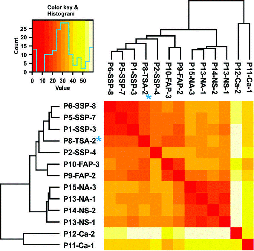
- Dehghanizadeh, S.; et al. Active BRAF-V600E is the key player in the generation of a sessile serrated polyp-specific DNA methylation profile. PloS one. 2018. Distributed under Open Access license CC BY 4.0, without modification.
For Research Use Only.