Retinal Vein Occlusion Modeling & Pharmacodynamics Services
Introduction
Retinal vein occlusion stands as a major eye condition that serves as an indicator of increased cardiovascular disease risk and stroke mortality among older individuals. Exudates, capillary non-perfusion, collateral formation, and microaneurysms serve as the typical symptoms. Ischemic RVO often complicates with macular edema (ME) and the formation of retinal and iris neovascularization, leading to severe vision loss. RVO is classified into central RVO (CRVO) and branch RVO (BRVO). CRVO includes superficial and deep retinal hemorrhages (HEs), which are dispersed around the veins near the lamina cribrosa, while BRVO involves hemorrhage in small veins from the retina to the supplied area caused by arterial compression of the vein. The most commonly used animals for establishing RVO animal models are non-human primates, followed by rodents and pigs. Creative Biolabs simulates the occurrence and development of retinal vein occlusion (RVO) through animal models, studying its risk factors and pathogenesis to promote drug development and treatment optimization for this disease.
Laser-Induced Retinal Vein Occlusion (RVO) Models
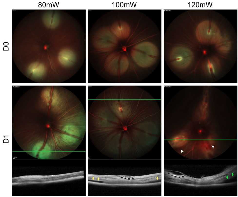 Fig. 1 Typical fundus changes of 80mW, 100mW, and 120mW groups after RVO.1
The leading cause of vision loss in older adults is diabetic retinopathy followed by RVO. The induction of RVO in mice through intravenous photosensitizer administration followed by laser photocoagulation remains a standard approach. The laser activates the photosensitizer at its peak absorption wavelength which generates singlet oxygen to damage the venous endothelium and initiates platelet adhesion and aggregation resulting in photothrombosis formation.
In reported mouse RVO models, the laser energy required to block the vein varies significantly, ranging from 100 mJ to over 3000 mJ. After intravenous injection of the photosensitizer rose bengal in mice, anesthesia is administered immediately. A coverslip coated with a viscoelastic agent is used to flatten the cornea, ensuring the visibility of the fundus structures, and laser surgery is performed once the surgical anesthesia state is achieved. The most representative pathologies of the RVO model include retinal swelling and thickening, accompanied by the formation of dense retinal cysts, as well as the development of intraretinal hemorrhages and exudative retinal detachment. Research has shown that after intravenously injecting rose bengal into cynomolgus monkeys and using laser exposure to induce RVO, the method is like that in mice. Retinal edema and vascular leakage along with foveal thinning were the observed pathological changes after modeling. The aqueous humor exhibits substantial upregulation of VEGF along with interleukin-6 and monocyte chemotactic protein-1. Animal disease models help decrease drug testing duration and expenses which facilitates quicker movement from drug development into clinical use.
Measurements
Creative Biolabs offers detection services for retinal vein occlusion models, including:
- Optical Coherence Tomography (OCT)
- Immunohistochemistry (TNF-α, VEGF)
- Spectral-domain optical coherence tomography (SD-OCT)
- Fluorescein angiography (FA)
- Scanning laser ophthalmoscopy (SLO)
Related Ocular Disease Models
In addition, other examples of ocular disease models available include:
- Dry Eye Models
- Corneal Disease Models
- Cataract Models
- Glaucoma Models
- Dry Age-Related Macular Degeneration (AMD) Models
- Wet Age-Related Macular Degeneration (AMD) Models
- Fundus Disease Models
- Diabetic Retinopathy Models
- Retinal Fibrosis Models
- Ocular Inflammation Models
Creative Biolabs has developed a thorough evaluation platform for disease animal models. For inquiries, please refer to the contact details provided on the company's homepage.
Reference
- Xu, Xiaowei, et al. "Exploring laser-induced acute and chronic retinal vein occlusion mouse models: Development, temporal in vivo imaging, and application perspectives." Plos one 19.6 (2024): e0305741. Distributed under Open Access license CC BY 4.0, without modification.
For Research Use Only.