
Duchenne Muscular Dystrophy (DMD) Modeling & Pharmacodynamics Service
Introduction
Duchenne Muscular Dystrophy (DMD) is a fatal, X-linked recessive neuromuscular disorder and the most prevalent form of muscular dystrophy in children. Caused by mutations in the DMD gene, the disease results in a complete absence of the dystrophin protein, a critical structural component that stabilizes the sarcolemma during muscle contraction. Without dystrophin, muscle fibers undergo chronic waves of degeneration and regeneration, triggering the accumulation of inflammatory cells, extensive fibrosis, and the eventual replacement of functional muscle with fatty tissue. Clinically, this pathology manifests in early childhood with delayed motor milestones and difficulty in standing (Gower's sign). The disease is characterized by rapidly progressive muscle wasting, typically leading to wheelchair dependence by age 12 and premature death, often due to respiratory failure or progressive cardiomyopathy, before the third decade. Current therapeutic approaches remain largely palliative, relying on glucocorticoids to manage symptoms. Creative Biolabs' DMD platform addresses the urgent need for curative interventions by providing high-fidelity animal models and multidimensional evaluation services. By simulating the complex interplay of muscle disruption, chronic inflammation, and fibrotic accumulation, we empower researchers to accelerate the translation of novel gene therapies, exon-skipping oligonucleotides, and small-molecule candidates from bench to bedside.
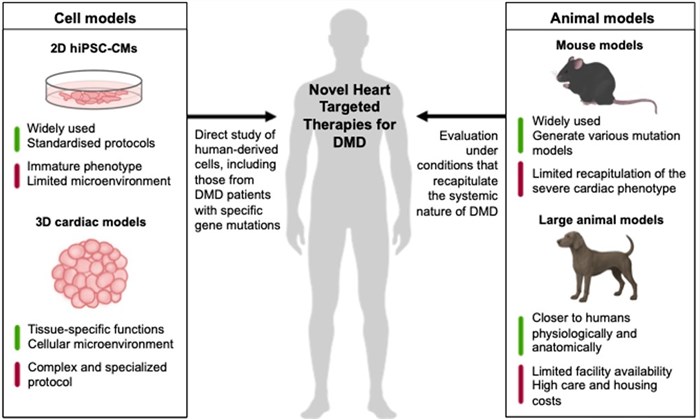 Fig.1 Cell and animal cardiac models for developing novel heart-targeted therapies for DMD.1
Available Duchenne Muscular Dystrophy Model
To assess the therapeutic potential of interventions for Duchenne Muscular Dystrophy (DMD), we provide a specialized DMD Drug Discovery & Evaluation Platform. This suite utilizes high-fidelity genetic models, such as gene knockout (KO) and humanized models, to replicate the hallmark pathologies of the disease, allowing for the precise validation of cutting-edge genetic therapies and muscle-stabilizing compounds.
| DMD Model | Models | Application Values | Animal Species |
| Genetic Models | KO, Humanized models (e.g., Dmd, Sspn, Itga7) | Ideal for studying dystrophin deficiency, muscle fiber necrosis, and fibro-adipogenic progenitor (FAP) activity. Test for studying muscle regeneration and screening membrane-stabilizing drugs, validating exon-skipping therapies, AAV-delivered strategies, or antisense oligonucleotides (ASOs). | Mouse |
Evaluation Platform
To provide an all-encompassing assessment of therapeutic efficacy, our platform integrates molecular, histological, and functional readouts to quantify disease progression and therapeutic rescue across multiple scales:
- Muscle Histopathology & Immunofluorescence (IF): Quantitative analysis of Centrally Nucleated Fibers (CNF) and fiber size distribution via Hematoxylin and Eosin (H&E), evaluation of interstitial fibrosis via Sirius Red or Masson's Trichrome, and high-resolution IF for sarcolemmal localization of the Dystrophin-Associated Glycoprotein Complex (DAGC).
- Biochemical & Inflammatory Markers: High-sensitivity measurement of creatine kinase (CK) and lactate dehydrogenase (LDH) in serum to assess sarcolemmal integrity, alongside quantification of pro-fibrotic and inflammatory drivers like TGF-β and TNF-α in muscle homogenates.
- Functional & Motor Performance: Rigorous behavioral testing, including Grid Hanging, Rotarod, and Treadmill (Exhaustion Test), to establish a direct correlation between molecular rescue and improvements in global muscle strength and endurance.
- In Vivo Imaging: Non-invasive, longitudinal monitoring of muscle inflammation, edema, and fatty infiltration utilizing high-resolution Magnetic Resonance Imaging (MRI) or Micro-Computed Tomography (Micro-CT) to track therapeutic response in real-time.
- Advanced Molecular Analysis: Utilizing Western Blot and the automated Capillary Electrophoresis for the absolute quantification of dystrophin protein restoration and Droplet Digital PCR (ddPCR) or RT-qPCR to precisely measure exon-skipping efficiency and mRNA stability.
Applications
- Genetic & Nucleic Acid Therapies: Comprehensive evaluation of AAV-delivered micro-dystrophin biodistribution and Exon-Skipping Oligonucleotides (ASOs), focusing on reading frame restoration, mRNA stability, and protein expression levels.
- Anti-fibrotic & Anti-inflammatory Agents: Specialized testing of candidates targeting TGF-β signaling pathways or macrophage polarization (M1 to M2 shift) to mitigate chronic inflammation and muscle scarring.
- Membrane Stabilizers & Small Molecules: Rigorous screening of compounds designed to reinforce the Dystrophin-Glycoprotein Complex (DGC), enhancing sarcolemmal integrity and preventing contraction-induced damage.
- Lead Optimization & Durability Studies: Longitudinal assessments to determine the persistence of therapeutic effects and optimize dosing regimens for clinical translation.
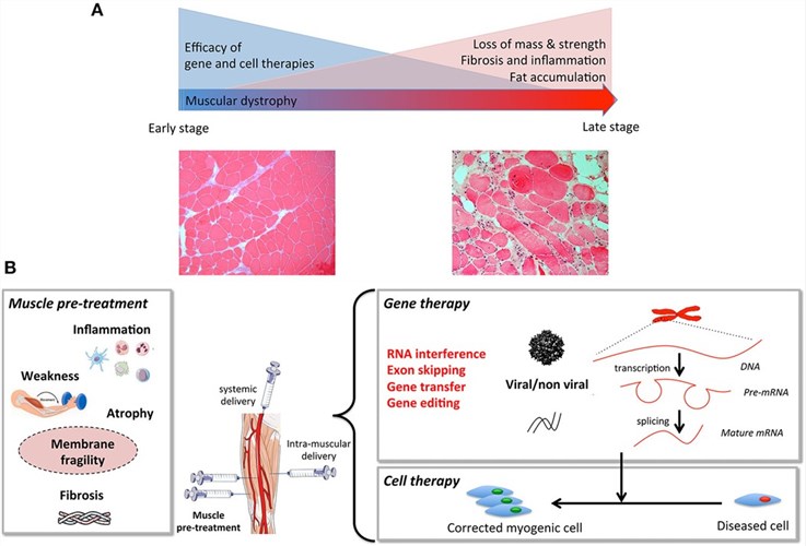 Fig.2 To optimize the efficacy of gene and cell therapies, integrated strategies must address progressive muscle degeneration, including mass loss, fibrosis, and fatty infiltration, by utilizing pre- or co-treatments that target inflammation, membrane fragility, and atrophy to prime the muscle tissue.2
Our Advantages
- Technical Accuracy: Standardized SOPs for muscle force measurements to ensure low coefficient of variation (CV).
- High-Fidelity Replication: Access to severe models that better predict clinical outcomes in ambulatory and respiratory function.
- Translational Expertise: Capabilities to correlate histological "Dystrophin-positive fibers" with functional "Grip Strength" improvements.
- Standardized Breeding: Large-scale colonies of mdx and humanized models to ensure immediate project initiation.
Work with Us
- Summarize the project requirements and fill in the information collection form.
- Sign a CDA from both parties to further communicate information, such as targets.
- Select an animal model, discuss experimental design, and determine assay parameters.
- Project costing and project schedule forecasting.
- We provide a detailed project plan, including the required sample quantities, methods, and protocols.
- Both parties confirm the project details and start the project.
- Confirm the timeline of the project.
- We provide periodic results and information on the animal's condition.
- We will work together to make project adjustments as necessary.
- We provide a comprehensive project report promptly.
- We arrange transportation for the produced samples.
- We provide a discussion of the project results and help to arrange the next steps.
- Data storage and archiving.
FAQs
-
Q: Which DMD mouse models are most suitable for gene therapy validation?
A: For AAV-micro-dystrophin or ASO exon-skipping therapies, we recommend hDMD (humanized) or Dmd-KO mice. The hDMD model is specifically required for testing human-sequence-specific RNA therapeutics to ensure accurate reading frame restoration.
-
Q: At what age should treatment begin in the mdx model?
A: For prevention studies, we typically start at 3-4 weeks (post-weaning). For "rescue" studies focusing on established pathology, we start at 8-12 weeks.
-
Q: Can you measure cardiac involvement in DMD models?
A: Yes, we offer echocardiography (Echo) and Electrocardiogram (ECG) to monitor cardiomyopathy, which typically develops in older mdx mice or early in double-knockout models.
-
Q: How do you verify AAV delivery to the diaphragm?
A: We perform specialized dissection and IF or Western Blot analysis specifically for the diaphragm, as it is a critical muscle for respiratory-related DMD outcomes.
-
Q: What is the typical study duration for a lead optimization program?
A: While acute markers can be seen in weeks, most DMD efficacy studies range from 8 to 24 weeks to sufficiently observe histopathological remodeling, functional improvements, and the persistence of gene expression.
Published Data
Intravenous delivery of Wharton's Jelly-derived Mesenchymal Stem Cells (WJ-MSCs) significantly attenuates fibrosis in the skeletal muscles and diaphragm of DMD mouse models. The observed anti-fibrotic efficacy is both regionally extensive and mechanistically consistent, establishing a robust preclinical foundation for the clinical translation of WJ-MSCs in DMD therapy.
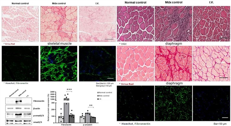 Fig. 2 Anti-fibrotic effect of WJ-MSCs in the skeletal muscle and diaphragm of mdx mice.3
References
- Moriyama, Hidenori, and Toshifumi Yokota. "Cardiac Cell and Animal Models for Duchenne Muscular Dystrophy in the Era of Gene Therapy and Precision Medicine." Cells vol. 14,17 1326. Distributed under Open Access license CC BY 4.0, without modification. https://doi.org/10.3390/cells14171326
- Cordova, Gonzalo et al. "Combined Therapies for Duchenne Muscular Dystrophy to Optimize Treatment Efficacy." Frontiers in Genetics vol. 9 114. Distributed under Open Access license CC BY 4.0, without modification. https://doi.org/10.3389/fgene.2018.00114
- Park, Sang Eon et al. "Wharton's Jelly-Derived Mesenchymal Stem Cells Reduce Fibrosis in a Mouse Model of Duchenne Muscular Dystrophy by Upregulating microRNA 499." Biomedicines vol. 9,9 1089. Distributed under Open Access license CC BY 4.0, with modification. https://doi.org/10.3390/biomedicines9091089
For Research Use Only.