Dry Eye Modeling & Pharmacodynamics Services
Introduction
Dry eye is a multifactorial tear and ocular surface disease characterized by alterations in tear secretion and quality, accompanied by discomfort and visual impairment such as dryness, redness, and foreign body sensation. Animal experimentation serves as an important adjunct to dry eye research, with the establishment of a suitable model being a prerequisite for study. Currently, various methods are available for establishing animal models of dry eye, including environmental factors, toxic substances, drug effects, and changes in sex hormone secretion levels. Creative Biolabs has developed a range of proven dry eye rodent models to help our customers more effectively explore the mechanisms of dry eye syndrome and develop research programs for efficacy.
Dry Eye Models Available at Creative Biolabs
- Scopolamine-Induced Dry Eye Models
Scopolamine is an anticholinergic drug with strong peripheral effects and can be used as a parasympathetic nerve inhibitor. In the study of dry eye modeling, its role is to block the cholinergic receptor of the lacrimal gland and reduce the secretion of tears. The model is like the clinical manifestations and pathogenesis of DED. After subcutaneous injection of scopolamine in rats, the lacrimal gland secretion was significantly reduced, including water, electrolytes, and protein.
- Benzalkonium Chloride-Induced Dry Eye Models
Benzalkonium chloride is the most commonly used preservative in eye drops. It can destroy the tear film, damage the corneal and conjunctival epithelium and intercellular junction proteins, damage the secretion function of goblet cells, induce ocular surface inflammation, decrease the stability of the tear film, and cause dry eye symptoms. The dry eye model of mice can be established by the topical application of BAC solution to the eyes of mice for 7 days.
- Environmental Stress-Induced Dry Eye Models
This model simulates evaporative dry eye by controlling external environmental conditions such as ventilation and humidity. The mice are exposed to a fan for 5 hours per day in a cubicle for 3 days. The external environment significantly decreased tear volume and increased corneal fluorescein and lissamine green staining scores. In addition, the corneal subbasal nerve density is severely damaged after exposure.
- Sleep Deprivation-Induced Mouse Dry Eye Model
A sleep deprivation (SD) mouse model is established by using the method of 'stick over water' to observe the effect of insufficient sleep on ocular surface health. When the mice stand on the stick and fall asleep, they fall into the water and wake up quickly. Mice are subjected to sleep disruption for 15-20 hours a day for a total of 1300 hours. The results show that sleep deprivation could reduce tear secretion, leading to corneal epithelial cell defects, and corneal sensitivity.
- Low Humidity Environment and Cholinergic Receptor Antagonists-Induced Dry Eye Models
Parasympathetic nerve excitation can increase tear secretion. Cholinergic receptor blockers such as scopolamine can significantly reduce tear secretion by inhibiting parasympathetic nerve excitation. A dry environment with low humidity can well simulate the long-term dry state of the human eye. The mixed dry eye model was stably established by scopolamine combined with a dehumidification drying device.
Measurements
At Creative Biolabs, our professional team has years of experience utilizing ophthalmic animal models for drug development. We also have extensive experience in various routes of administration and can customize studies depending on your needs. In addition, we conduct a full range of measurements to ensure the modeling outcomes, as well as investigating drug efficacy, including but not limited to:
- Schirmer test (SIT)
- Break-up time (BUT)
- Fluorescein staining (FLS)
- Real-time PCR (e.g. TNF-α, IFN-γ, IL-1β)
- Histopathological observation (e.g., cornea, conjunctiva, lacrimal gland, Hartman's gland)
Related Ocular Disease Models
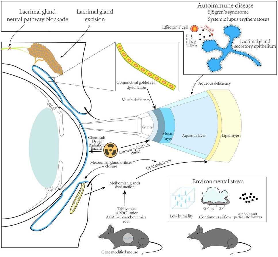 Fig. 1 The principle of animal dry eye modeling.
This schematic diagram shows the method of developing animal dry eye models.1
Except for the dry eye models, we can also provide the following other ocular disease models to our global customers.
- Corneal Disease Models
- Cataract Models
- Glaucoma Models
- Dry Age-Related Macular Degeneration (AMD) Models
- Wet Age-Related Macular Degeneration (AMD) Models
- Fundus Disease Models
- Diabetic Retinopathy Models
- Retinal Fibrosis Models
- Retinal Vein Occlusion Models
- Ocular Inflammation Models
Creative Biolabs has an animal efficacy evaluation model with good evaluation ability, which helps with the clinical transformation of new drugs. You can find our contact information on the company's home page.
Reference
- Zhu, Jun, et al. "Application of animal models in interpreting dry eye disease." Frontiers in Medicine 9 (2022): 830592. Distributed under Open Access license CC BY 4.0, without modification.
For Research Use Only.