Dry Age related Macular Degeneration (AMD) Modeling & Pharmacodynamics Services
Introduction
Age-related macular degeneration (AMD) is a degenerative disease that occurs in the macular area. It is one of the main causes of blindness in people over 50 years old worldwide. There are two main types of AMD, neovascular AMD ("wet" AMD) and non-neovascular AMD ("dry" AMD). Dry AMD accounts for about 80% to 85% of all cases and usually has a good visual prognosis. The pathological changes of dry AMD include degenerative changes in photoreceptor cells and retinal pigment epithelium (RPE) cells, accumulation of lipofuscin, and drusen formation. Anti-vascular endothelial growth factor has achieved significant clinical efficacy in the treatment of wet AMD, but there is no effective treatment for dry AMD. Appropriate animal models are essential for the study of dry AMD and contribute to drug development. Creative Biolabs uses animal models to simulate the onset and progression of AMD, studying its risk factors and pathogenesis to promote the development of drugs and the improvement of treatment methods for this condition.
Dry AMD Disease Models Available at Creative Biolabs
- Sodium Iodate (NaIO3)-Induced Retinal Degeneration
NaIO3 is widely used to simulate AMD because it can easily and quickly induce retinal oxidative stress, resulting in RPE and photoreceptor death within one week after injection. After NaIO3 was injected into the orbital venous plexus of mice, the ERG waveform decreased significantly. Histological sections showed that the RPE layer had pigment disorder, and the retinal outer nuclear layer gradually became thinner with the extension of injection time.
- Blue/White Light-Induced Retinal Degeneration
Light can induce photoreceptor molecules, such as rhodopsin and lipofuscin, to produce reactive oxygen species, resulting in lipid peroxidation of the outer segment disc of photoreceptor cells, thereby inducing apoptosis of photoreceptor cells and causing damage to the retina. Blue light exposure immediately led to vacuolization of retinal pigment epithelial cells and swelling of the outer nuclear layer and photoreceptor inner segments.
- N-methyl-N-nitrosourea (MNU)-Induced Retinal Degeneration
MNU is a strong carcinogen, teratogen, and mutagen in experimental animals and is commonly used in animal models to chemically induce photoreceptor degeneration. After intraperitoneal injection of MNU in C57/BL mice, the thickness of the outer nuclear layer (ONL) gradually decreased, which was related to the loss of the outer segment of the photoreceptor, bipolar cell dendritic retraction, and reactive gliosis.
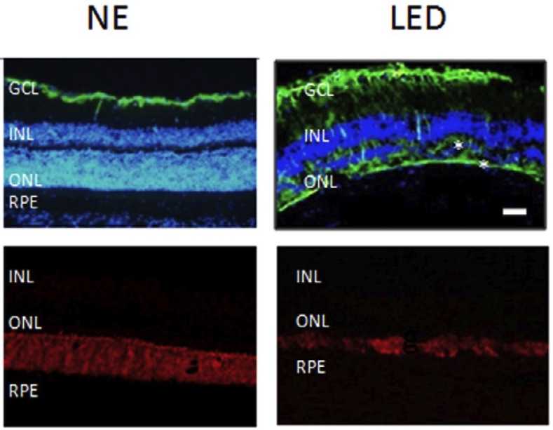 Fig. 1 Images of retinal damage caused by LED exposure in rats.1
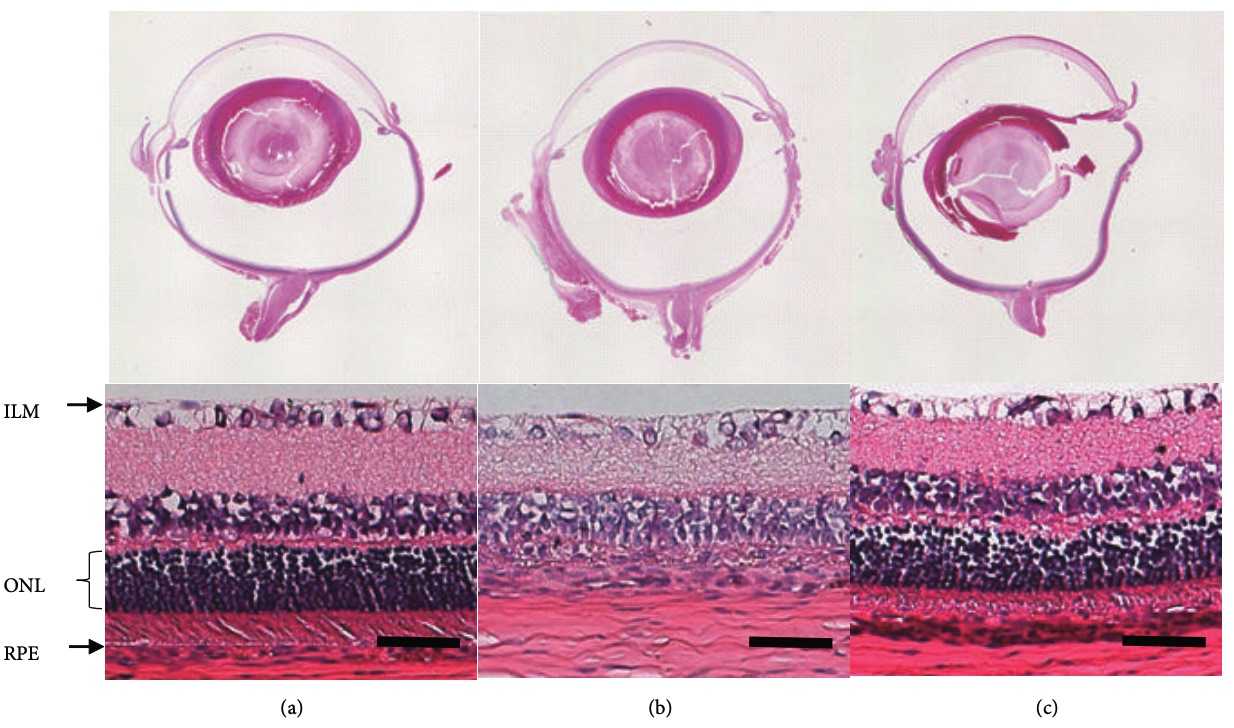 Fig. 2 Histological retinal images of MNU injected rats.2
Measurements
Creative Biolabs provides detection services of dry age-related macular degeneration models, such as:
- Electroretinograms (ERG)
- Transmission electron microscopy (TEM)
- Optical Coherence Tomography
- Western Blot Analysis (JNK, p38, PARP and AIF)
- Immunohistochemistry (PNA, GFAP, IBA1, ED1, GS, pERK)
Related Ocular Disease Models
Except for the dry AMD models, we can also provide the following other ocular disease models to our global customers.
- Dry Eye Models
- Corneal Disease Models
- Cataract Models
- Glaucoma Models
- Wet Age-Related Macular Degeneration (AMD) Models
- Fundus Disease Models
- Diabetic Retinopathy Models
- Retinal Fibrosis Models
- Retinal Vein Occlusion Models
- Ocular Inflammation Models
Creative Biolabs has established a comprehensive evaluation system for disease animal models. Please feel free to contact us for more information.
References
- Krigel, Arthur, et al. "Light-induced retinal damage using different light sources, protocols and rat strains reveals LED phototoxicity." Neuroscience 339 (2016): 296-307. Distributed under Open Access license CC BY 4.0. The image was modified by extracting and using part of the original image.
- Sugano, Eriko, et al. "N‐Methyl‐N‐Nitrosourea‐Induced Photoreceptor Degeneration Is Inhibited by Nicotinamide via the Blockade of Upstream Events before the Phosphorylation of Signalling Proteins." BioMed Research International 2019.1 (2019): 3238719. Distributed under Open Access license CC BY 4.0. The image was modified by extracting and using a-c part of the original image.
For Research Use Only.