Fundus Disease Modeling & Pharmacodynamics Services
Introduction
Fundus diseases encompass various conditions that target the eye's posterior parts such as the retina, choroid, and optic nerve. Visual function becomes impaired by these disorders which can result in complete blindness in severe cases. The primary categories of this condition include central retinal artery occlusion and central serous chorioretinopathy along with floaters. Animal models serve as indispensable tools for researching the origin and progression of eye diseases and developing diagnostic and treatment strategies. Researchers use mice, rats, rabbits, pigs, and primates as animal models to study fundus diseases. Creative Biolabs uses animal models to recreate the development of fundus disease and investigates its risk factors and pathogenesis to advance drug development and treatment approaches.
Fundus Disease Models Available at Creative Biolabs
- Hyperoxia-Induced Retinal Neovascularization Models
Hyperoxia-induced retinal neovascularization models serve as a traditional animal model for researching abnormal retinal vascular growth and related diseases like retinopathy of prematurity and diabetic retinopathy. Researchers apply high-oxygen conditions to experimental animals for a certain period to disturb normal retinal vascular growth before returning them to normal oxygen levels which results in pathological neovascularization. The model measures drug effects through quantitative analysis of avascular regions and neovascular volume alongside immunohistochemistry detection of pivotal molecules.
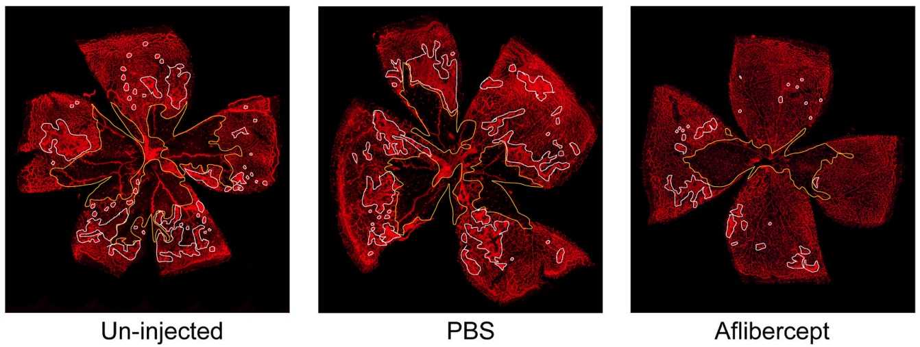 Fig. 1 Representative images of flat-mounted retinas of hyperoxia-induced retinal neovascularization models in mice.1 The avascular area is delineated in yellow, and the area of pre-retinal neovascularization is delineated in white.
- High intraocular pressure (IOP)-Induced Retinal Ischemia-Reperfusion Injury Models
The IOP-induced retinal ischemia-reperfusion injury model serves as an experimental system to investigate acute glaucoma alongside retinal vascular disorders. Artificial elevation of intraocular pressure through saline or air infusion produces ischemia by obstructing blood flow above retinal perfusion pressure which then leads to reperfusion when blood flow returns to normal. The ischemic phase disrupts retinal energy metabolism which results in the destruction of cells through either apoptosis or necrosis. The reperfusion phase causes damage to increase because of the excessive production of reactive oxygen species (ROS), calcium overload, and the release of inflammatory factors like TNF-α and IL-6 which trigger oxidative stress and inflammatory responses. Researchers benefit from this model as it enables exact experimental condition management while achieving fast induction and consistent results that mirror acute high IOP-induced pathological damage. The investigation of I/R injury mechanisms and therapeutic outcomes is possible through histological analysis as well as functional tests like ERG and molecular studies. The model serves as an important tool for neuroprotective agent screening and antioxidant and anti-inflammatory therapy development in acute glaucoma and retinal vascular disease research.
Measurements
Creative Biolabs can provide a variety of testing parameters for fundus disease models, such as:
- Transmission electron microscopy (TEM)
- Optical Coherence Tomography (OCT)
- Fluorescein angiography (FA)
- Scanning laser ophthalmoscopy (SLO)
Related Ocular Disease Models
Except for fundus disease models, we can also provide the following other ocular disease models to our global customers.
- Dry Eye Models
- Corneal Disease Models
- Cataract Models
- Glaucoma Models
- Dry Age-Related Macular Degeneration (AMD) Models
- Wet Age-Related Macular Degeneration (AMD) Models
- Diabetic Retinopathy Models
- Retinal Fibrosis Models
- Retinal Vein Occlusion Models
- Ocular Inflammation Models
Creative Biolabs' experienced scientists have established a complete preclinical efficacy evaluation platform and a comprehensive disease animal model evaluation system. We can provide customers with professional and customized services. If you need, please feel free to contact us.
Reference
- Klaska, Izabela P., et al. "Intravitreal administration of recombinant human opticin protects against hyperoxia-induced pre-retinal neovascularization." Experimental eye research 215 (2022): 108908. Distributed under Open Access license CC BY 4.0. The image was modified by extracting and using the E part of the original image.
For Research Use Only.