
Huntington's Disease (HD) Modeling & Pharmacodynamics Service
Introduction
Huntington's Disease (HD) is a devastating, autosomal dominant neurodegenerative disorder caused by a cytosine-adenine-guanine (CAG) trinucleotide repeat expansion in the huntingtin (HTT) gene. Affecting approximately 5 to 10 per 100,000 individuals in Western populations. At the structural level, the disease manifests as profound atrophy within the basal ganglia; widespread morphological changes in the caudate and putamen are observed, with significantly greater tissue deflation in pre-symptomatic (pro-HD) individuals compared to healthy controls. This genetic defect leads to the selective and progressive loss of GABAergic medium spiny neurons (MSNs) in the striatum. To address this complexity, Creative Biolabs provides a specialized bridge for HD drug development, combining genetically precise animal models with advanced neurobehavioral and molecular readouts to rigorously evaluate huntingtin-lowering therapies and neuroprotective agents.
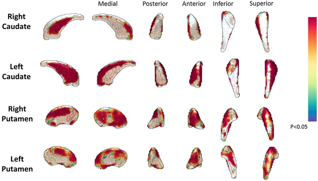 Fig.1 Vertex-wise shape differences between individuals with prodromal HD and healthy controls. The colour bar represents the percentage of atrophy at a specific vertex in the disease group relative to the control group.1
Available Huntington's Disease Models
We offer a diverse portfolio of models ranging from rapid-screening transgenic fragments to slow-progressing, translationally relevant knock-ins. This allows us to align the model's kinetics with your drug's specific mechanism of action.
| HD Models | Modeling Methods | Application Values | Animal Species |
| Quinolinate-Induced HD Model | Intrastriatal microinjection of NMDA receptor agonist (Quinolinic acid). | Striatal Atrophy & Cell Therapy Validation: Replicates selective GABAergic neuronal loss. Specifically designed for evaluating NMDA receptor antagonists, stem cell transplantation, and cell-replacement therapies. | Mouse |
| 3-NY-Induced HD Model | Systemic administration of 3-Nitropropionic acid (mitochondrial toxin). | Bioenergetic Profiling & Mechanism Elucidation: Precisely mimics the metabolic dysfunction and oxidative stress hallmarks of HD. Optimized for evaluating mitochondrial-targeted therapies, antioxidants, and metabolic regulators. | Mouse |
| R6/2 Mouse HD Model | Transgenic expression of human N-terminal HTT fragment (~150 CAG repeats). | Accelerated Efficacy & Survival Screening: Features a rapid and robust disease phenotype. Ideal for high-throughput screening of neuroprotective agents and therapies aimed at extending survival or alleviating motor symptoms. | Rat |
| Q175 Mouse HD Model | Human HTT exon 1 (~175 CAG repeats) knocked into the mouse Htt locus. | The Premier Platform for DMTs: Mimics chronic human disease progression. The gold standard for validating HTT-lowering (ASO/siRNA), gene editing, and long-term efficacy profiling. | Mouse |
Evaluation Platform
To provide a gold-standard assessment of HD therapeutics, we offer a comprehensive evaluation platform that tracks the disease's characteristic "triad" of symptoms: motor dysfunction, cognitive decline, and psychiatric disturbances, alongside state-of-the-art molecular quantification.
-
Behavioral Phenotyping
-
Motor Function & Coordination:
- Hind-limb Clasping: The primary neurological marker for HD in rodents; used to track the progression of striatal dysfunction.
- Rotarod & Beam Walk: Quantifying deficits in balance, gait fluidity, and fine motor coordination.
- Gait Analysis (Fine Motor Kinematics): Utilizing automated systems to detect subtle changes in stride length and paw pressure.
-
Cognitive & Executive Function:
- Novel Object Recognition (NOR): Assessing short-term recognition memory and interest in novelty.
- T-Maze / Y-Maze: Measuring spontaneous alternation and spatial working memory, which are often impaired early in HD.
-
Neuropsychiatric Profiling:
- Prepulse Inhibition (PPI): A critical assay for sensorimotor gating deficits, replicating the "startle" abnormalities seen in HD patients.
- Open Field Test: Monitoring anxiety-like behaviors and general locomotor activity (hyperactivity vs. hypoactivity).
-
Motor Function & Coordination:
-
Molecular PD & Biomarker Analysis
-
mHTT Quantification:
- Ultra-Sensitive Assays: Utilizing (Meso Scale Discovery) MSD to quantify mutant Huntingtin (mHTT) protein levels in the striatum, cortex, and CSF. This is the "gold standard" for validating ASO or siRNA efficacy.
- mRNA Expression: Quantitative RT-PCR to verify gene knockdown at the transcriptional level.
-
Translational Biomarkers:
- Neurofilament Light Chain (NfL): High-sensitivity detection of NfL in plasma or CSF as a dynamic indicator of axonal degeneration and treatment response.
-
mHTT Quantification:
-
Quantitative Histopathology & Imaging
-
Striatal Integrity:
- DARPP-32+ Neuron Counting: Stereological quantification of medium spiny neurons (MSNs) to assess the prevention of striatal atrophy.
- Striatal Volume: Volumetric analysis of the striatum and ventricles via high-resolution MRI or histological reconstruction.
-
Proteinopathy Analysis:
- Inclusion Body Quantification: Using EM-48 or S830 antibodies to visualize and count nuclear and cytoplasmic mHTT aggregates.
- Synaptic Integrity: Quantification of markers such as Synaptophysin or PSD-95 to evaluate the preservation of neural circuitry.
-
Striatal Integrity:
-
Advanced In Vivo Monitoring
- In Vivo Imaging: Structural MRI to monitor progressive brain atrophy and ventricular enlargement longitudinally in the same animal.
- Electrophysiology: Ex vivo brain slice recording to measure changes in synaptic plasticity (LTP/LTD) and MSN excitability.
Applications
- HTT-Lowering Therapies: Validating the potency of ASOs, siRNAs, and gene editing by quantifying the knockdown of mHTT mRNA and protein levels across target brain regions.
- Autophagy Modulators: Evaluating small molecules that enhance the clearance of mHTT aggregates to mitigate proteotoxic stress and promote neuronal rescue.
- Neuroprotective Screening: Assessing mitochondrial stabilizers, anti-apoptotic agents, and Sigma-1 agonists (e.g., Pridopidine) to preserve synaptic integrity and bioenergetic health.
- Biomarker Development: Correlating mHTT and NfL levels in the CSF/Plasma with motor functional improvements to align preclinical efficacy with clinical trial endpoints.
- Preclinical Efficacy & Drug Development: Delivering comprehensive PK/PD profiling and robust efficacy data in validated HD models to support lead optimization and IND-enabling studies.
Our Advantages
- Validated Genetic Stability: We perform routine fragment analysis to verify CAG repeat lengths, preventing "phenotype drift" and ensuring consistent disease onset across all study cohorts.
- Diverse Model Portfolio: Access to high-fidelity transgenic and knock-in lines, offering both mouse and rat versions to accommodate varied PK/PD requirements and surgical complexities.
- Spatial Precision: Expert stereotaxic delivery and micro-dissection techniques allow for targeted intervention and region-specific analysis of the striatum and cortex.
- mHTT Proteodynamics: Utilization of ultra-sensitive platforms, including MSD, TR-FRET, and SIMOA, to quantify mutant Huntingtin (mHTT) protein levels in brain tissue, CSF, and plasma.
- Clinical Endpoint Alignment: All models are validated using the Clasping Test and advanced motor kinematics, providing a functional bridge to human clinical symptoms.
- Temporal Tracking: Our platform supports longitudinal studies spanning several months, enabling the evaluation of true disease-modifying potential rather than just temporary symptomatic relief.
- Multidimensional Data Integration: Correlation of behavioral recovery with structural preservation (via stereological neuron counting) and molecular target engagement for a comprehensive PD profile.
Work with Us
- Summarize the project requirements and fill in the information collection form.
- Sign a CDA from both parties to further communicate information, such as targets.
- Select an animal model, discuss experimental design, and determine assay parameters.
- Project costing and project schedule forecasting.
- We provide a detailed project plan, including the required sample quantities, methods, and protocols.
- Both parties confirm the project details and start the project.
- Confirm the timeline of the project.
- We provide periodic results and information on the animal's condition.
- We will work together to make project adjustments as necessary.
- We provide a comprehensive project report promptly.
- We arrange transportation for the produced samples.
- We provide a discussion of the project results and help to arrange the next steps.
- Data storage and archiving.
FAQs
-
Q: How do you ensure the stability of CAG repeats in your transgenic and knock-in models?
A: CAG repeat instability is a common challenge in HD research. We perform rigorous genotypic verification and fragment analysis on every animal used in a study to ensure the repeat length remains within the targeted range (e.g., ~150 for R6/2 or ~175 for Q175). This ensures cohort consistency and the reproducibility of the disease phenotype.
-
Q: Which model is best for testing Huntingtin (HTT) lowering therapies like ASOs or siRNA?
A: The Q175 Knock-In model is the preferred choice for HTT-lowering strategies. Unlike fragment models (like R6/2), the Q175 model contains the human HTT exon 1 within the native mouse Htt locus, allowing for the study of gene-silencing or gene-editing tools in a slow-progressing, translationally relevant context.
-
Q: What is the most sensitive method you use to quantify mutant Huntingtin (mHTT) protein?
A: We primarily utilize the MSD and TR-FRET platforms. These ultra-sensitive assays allow us to detect and quantify mHTT levels in small volumes of CSF, plasma, or specific brain regions (like the striatum and cortex) with high dynamic range.
-
Q: Can your platform distinguish between drug effects on mutant vs. wild-type Huntingtin?
A: Yes. We offer allele-specific RT-qPCR and protein assays that can distinguish between the mutant (mHTT) and wild-type (wtHTT) alleles. This is critical for evaluating the safety and selectivity of therapies designed to spare the essential functions of normal huntingtin.
-
Q: Why is the Clasping Test used instead of just Rotarod?
A: While the Rotarod measures general motor coordination, the Hind-limb Clasping reflex is a specific marker of neurological impairment and striatal dysfunction in HD mice. It mimics the dystonic postures seen in HD patients and often appears earlier and more consistently than gross motor failure.
-
Q: Do you offer longitudinal brain imaging for HD studies?
A: Yes. We can perform longitudinal MRI to monitor progressive striatal atrophy and ventricular enlargement in the same animal over several months. This provides powerful structural evidence of neuroprotection that correlates with our behavioral and histological findings.
-
Q: How do you quantify neurodegeneration in the striatum?
A: We use unbiased stereological counting of DARPP-32+ neurons. DARPP-32 is a highly specific marker for the Medium Spiny Neurons (MSNs) that are selectively lost in HD. Quantifying the preservation of these cells is the gold-standard histological endpoint for neuroprotective efficacy.
Published Data
Olaparib significantly attenuates neuroinflammation in the striatum of R6/2 HD mice by reducing GFAP+ astrogliosis and suppressing microglial hyperactivity. Notably, it promotes the morphological transition of microglia from an activated amoeboid state back to a quiescent ramified state, effectively restoring the homeostatic neural environment.
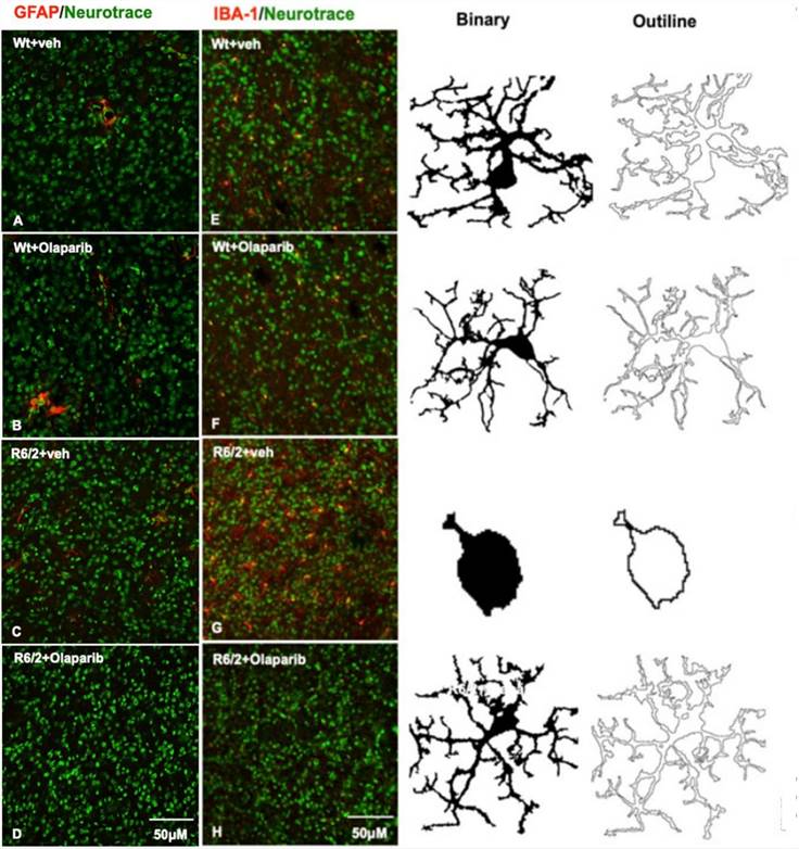 Fig. 2 Olaparib reduces astrogliosis and microglial activation.2
References
- Cruickshank, Travis et al. "Rate of torque development and striatal shape in individuals with prodromal Huntington's disease." Scientific Reports vol. 10,1 15103. Distributed under Open Access license CC BY 4.0, without modification. https://doi.org/10.1038/s41598-020-72042-2
- Paldino, Emanuela et al. "Modulation of Inflammasome and Pyroptosis by Olaparib, a PARP-1 Inhibitor, in the R6/2 Mouse Model of Huntington's Disease." Cells vol. 9,10 2286. Distributed under Open Access license CC BY 4.0, with modification. https://doi.org/10.3390/cells9102286
For Research Use Only.