Ocular Inflammation Modeling & Pharmacodynamics Services
Introduction
Ocular inflammatory diseases such as uveitis, bacterial conjunctivitis, and infectious keratitis create complex treatment dilemmas while threatening to cause vision loss and complete blindness. Researchers developed targeted and secure treatments for ocular inflammation and immune-mediated disorders because corticosteroids and nonsteroidal anti-inflammatory drugs used for eye inflammation produced damaging visual side effects. New ocular inflammation treatments require proper immune-related animal models to assess their safety and effectiveness. Creative Biolabs produces animal models that replicate retinal vein occlusion (RVO) development to study its risk factors and pathogenesis thus aiding drug development and treatment advancement.
Ocular Inflammation Models Available at Creative Biolabs
- Experimental Autoimmune Uveitis (EAU) Models
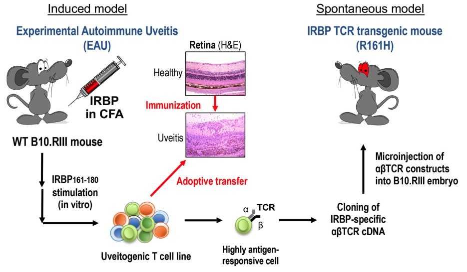 Fig. 1 Induced and spontaneous models of autoimmune uveitis.1
Autoimmune uveitis develops as a chronic inflammatory condition where the body's immune system targets and damages the uvea which constitutes the eye's middle layer. Patients with this condition frequently exhibit related autoimmune disorders and face a risk of vision impairment or total blindness. AU presents clinically through eye pain and blurred vision, redness while severe instances can damage essential eye parts including the retina and optic nerve. Experimental autoimmune uveitis (EAU) serves as a crucial animal model for research into the pathogenesis and immune responses of AU and for developing therapeutic approaches. The development of EAU occurs when retinal antigens stimulate immune responses in mice or rats, producing inflammation similar to the condition of AU. Through this model, scientists have obtained a better understanding of how T cells contribute to the inflammatory mechanisms in uveitis and discovered methods to modulate immune responses to prevent or treat the disease.
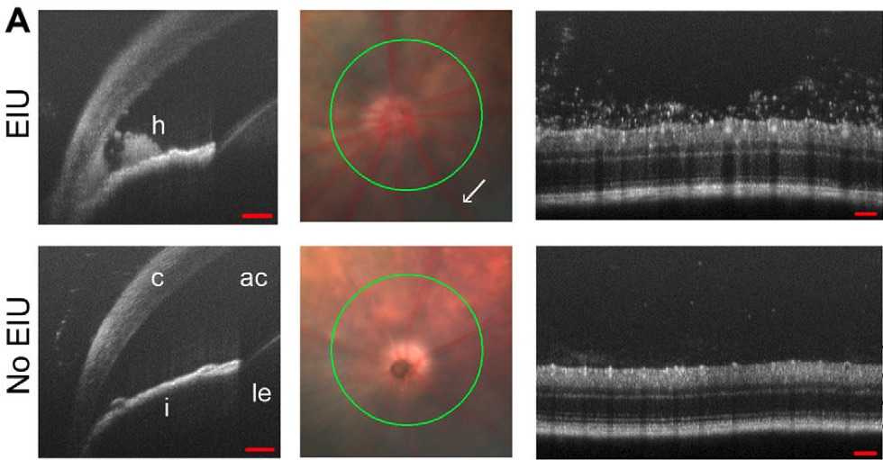 Fig. 2 Circular scans (green) were centred on the optic disc using
fundus images.2
Uveitis represents an inflammation of the uvea, which may result in vision loss. The disease maintains connections to numerous pathological conditions, which include autoimmune disorders like rheumatoid arthritis and systemic lupus erythematosus, along with bacterial and viral infections and chemical irritants as well as metabolic abnormalities. The disease pathology emerges from complex mechanisms that involve multiple immune system responses. Scientists commonly employ rodent models consisting of rats and mice to study endotoxin-induced uveitis (EIU) in their experimental research. The experimental procedure causes acute intraocular inflammation in animals by delivering bacterial endotoxins including lipopolysaccharide (LPS). The EIU model serves as a primary tool for scientists who study the inflammatory mechanisms of uveitis and evaluate potential anti-inflammatory medications. The EIU model demonstrates uveitis by producing inflammatory exudates and leukocyte infiltration inside both the anterior chamber and vitreous cavity. The brief nature of the inflammatory response observed renders this model ideal for studying inflammation's early stages.
- Graves' ophthalmopathy Models
Thyroid dysfunction causes Graves' Orbitopathy (GO), which is an autoimmune eye disorder that doctors call Thyroid Eye Disease (TED) and Thyroid-Associated Ophthalmopathy (TAO). Graves' Orbitopathy initiates localized inflammation and tissue swelling in orbital fibroblasts accompanied by adipose tissue growth that leads to fibrosis resulting in exophthalmos (bulging eyes), diplopia (double vision), and vision loss. The precise mechanism behind GO remains unclear but studies indicate that TSHR and TRAbs are critical factors in disease advancement. Thyroid-Stimulating Immunoglobulin (TSI) serves as a distinct biomarker that shows a strong correlation with both Graves' disease and GO clinical manifestations. Researchers create current animal models of GO using direct immunization as well as genetic immunization, cellular immunization, and drug induction techniques together with protein immunization. Evaluations based on ocular appearance assessments alongside serological tests and imaging studies plus pathological examinations offer a crucial understanding of GO mechanisms and assist in developing therapeutic strategies.
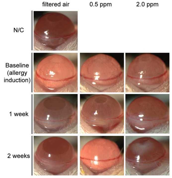 Fig. 3 Changes in conjunctival chemosis and conjunctival injection after exposure
to ozone in a mouse model of experimental allergic conjunctivitis.3
Allergic conjunctivitis represents an inflammation of the conjunctiva, which occurs when the immune system responds to antigens like pollen and animal dander among other environmental allergens. The condition manifests through multiple symptoms like eye itching, as well as hyperemia and edema. Researchers prefer the ovalbumin (OVA)-induced model to study allergic conjunctivitis since OVA serves as a standard antigen in allergy research. The reaction between antigens and antibodies triggered by OVA demonstrates the clinical signs of allergic conjunctivitis.
- Bacterial Conjunctivitis Models
Bacterial conjunctivitis stands as a common eye infection where Staphylococcus species along with Streptococcus and Haemophilus species usually participate. Eye redness and increased discharge characterize the bacterial infection while patients also experience stinging sensations and discomfort. People with normal immune function often experience self-resolving bacterial conjunctivitis symptoms that diminish within a timeframe of several days to one week. Healthy rabbits serve as models in scientific research for the study of bacterial conjunctivitis. The rabbits receive a pre-prepared bacterial suspension in their eyes which leads to the development of a significant yellowish discharge together with severe conjunctival redness and corneal clouding as well as tightly shut eyelids after 24 to 48 hours. Pathological sections demonstrate extensive corneal swelling accompanied by clouding. The anterior third region of the stroma contains many neutrophils together with lymphocytes, fibroblasts, and necrotic tissues.
Infectious keratitis develops from viral, bacterial, fungal, yeast, or amoebic infections and produces symptoms such as pain and impaired vision along with photophobia, redness, and a gritty sensation. The primary risk factors for developing infectious keratitis are eye trauma along with extended use of contact lenses and chronic ocular disease combined with previous eye surgery and corticosteroid usage. Researchers study infectious keratitis using animal models that primarily focus on bacterial infections from Pseudomonas aeruginosa and Staphylococcus aureus. Genetically modified rodents serve as models that reveal bacterial mechanisms and host immune reactions which provide insights to design better treatments for corneal damage prevention.
- Capsaicin-induced Corneal Neuritis Models
The cornea stands out as one of the human body's most sensitive regions because it contains an extensive network of nerve fibers. The cornea detects external stimuli acutely because of its high nerve density. Corneal sensory nerves serve dual roles by detecting pain and dangerous stimuli and maintaining corneal tissue health through environmental regulation. The compound capsaicin (8-methyl-N-vanillyl-6-nonenamide) present in chili peppers triggers irritation by stimulating primary sensory nerve endings and depleting neuropeptides. This process is followed by prolonged sensory nerve inactivation: Studies show that treating rats with capsaicin during their neonatal period results in selective sensory nerve inactivation which causes peripheral sensory fiber regeneration to be delayed and incomplete throughout adulthood.
Measurements
Creative Biolabs is capable of delivering detection services for various ocular inflammation models, such as:
- Color fundus photography
- Transmission electron microscopy (TEM)
- Optical Coherence Tomography (OCT)
- Fluorescein angiography (FA)
Related Ocular Disease Models
A further selection of ocular disease models is also offered, such as:
- Dry Eye Models
- Corneal Disease Models
- Cataract Models
- Glaucoma Models
- Dry Age-Related Macular Degeneration (AMD) Models
- Wet Age-Related Macular Degeneration (AMD) Models
- Fundus Disease Models
- Diabetic Retinopathy Models
- Retinal Fibrosis Models
- Retinal Vein Occlusion Models
Creative Biolabs has developed an extensive evaluation system for animal disease models. For more information, please refer to the contact details on the company's homepage.
References
- Horai, Reiko, and Rachel R. Caspi. "Microbiome and autoimmune uveitis." Frontiers in Immunology 10 (2019): 232. Distributed under Open Access license CC BY 4.0, without modification.
- Chu, Colin J., et al. "Multimodal analysis of ocular inflammation using the endotoxin-induced uveitis mouse model." Disease Models & Mechanisms 9.4 (2016): 473-481. Distributed under Open Access license CC BY 4.0. The image was modified by extracting and using A part of the original image.
- Lee, Hun, et al. "Effects of exposure to ozone on the ocular surface in an experimental model of allergic conjunctivitis." PLoS One 12.1 (2017): e0169209. Distributed under Open Access license CC BY 3.0. The image was modified by extracting and using part of the original image.
For Research Use Only.