
Orchiectomy induced Osteoporosis Modeling & Pharmacodynamics Service
Are you currently facing challenges such as inconsistent bone loss induction, high variability in preclinical data, or difficulty correlating biochemical markers with structural bone integrity? Our Orchiectomy induced Osteoporosis Modeling & Pharmacodynamics Service helps you obtain high-fidelity translational data and accelerate the development of male-specific bone therapeutics through our validated surgical protocols and multi-dimensional analysis platforms.
Overview of Orchiectomy induced Osteoporosis Modeling & Pharmacodynamics Service
The skeletal system is a dynamic tissue regulated by a delicate balance between osteoblastic bone formation and osteoclastic bone resorption. While osteoporosis is often categorized as a female health issue, male osteoporosis is a severe clinical condition frequently linked to primary hypogonadism or secondary causes such as androgen deprivation therapy (ADT) for prostate cancer. Orchiectomy (ORX) serves as the definitive surgical model to simulate this androgen-deficient state. By removing the primary source of testosterone, researchers can induce a high-turnover bone loss state that mimics the physiological deterioration of the male skeleton, characterized by reduced bone mineral density and compromised micro-architecture.
Orchiectomy induced Osteoporosis Model
Creative Biolabs provides a comprehensive suite of validated models to study bone metabolism. Our Orchiectomy (ORX) induced Osteoporosis Model specifically targets male bone loss pathways. We also offer the OVX induced Osteoporosis Model for estrogen-deficiency studies, the Glucocorticoid-Induced Osteoporosis Model for secondary osteoporosis research, and the Retinoic Acid-Induced Osteoporosis Model for rapid induction scenarios. These models utilize standardized surgical and pharmacological protocols in the rat and Rabbit species to ensure reproducible bone mass reduction and micro-architectural decay within predictable study timelines.
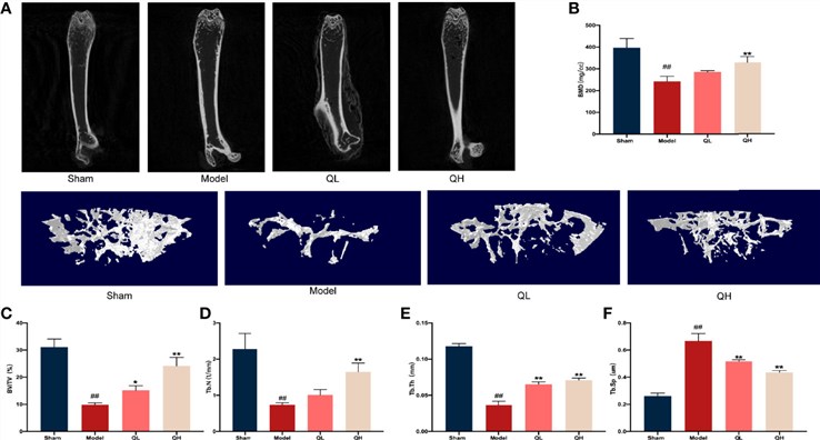 Fig.1 Quercetin-enhanced bone microstructure in orchiectomized mice.1
Evaluation Platform of Our Service
Creative Biolabs utilizes a multi-dimensional analysis platform to provide a holistic view of bone health and therapeutic efficacy. Our technical capabilities include:
-
Biochemical & Molecular Analysis
- Bone Turnover Markers: Serum/Urine assays for CTX-I, NTX-I, Osteocalcin, and ALP.
- Hormonal Profiling: Testosterone and Dihydrotestosterone (DHT) quantification.
- Molecular Expression: qPCR and Western Blotting for RANKL/OPG ratio and Sirt1 signaling.
-
Histopathological Examination
- Staining Protocols: H&E, Safranin-O/Fast Green, and Goldner's Trichrome.
- Dynamic Histomorphometry: Tetracycline/Calcein dual-labeling for Bone Formation Rate (BFR).
- Cellular Quantification: Osteoclast (TRAP-positive) and osteoblast counting.
-
Behavioral & Functional Testing
- Mechanical Testing: Three-point bending and femoral neck load-to-failure testing.
- Locomotor Assessment: Automated gait analysis and grip strength measurement.
-
Advanced Imaging & Instrumentation
- Micro-CT (High Resolution): Quantification of BV/TV, Tb.N, Tb.Th, and Tb.Sp.
- Dual-Energy X-ray Absorptiometry (DEXA): Longitudinal Bone Mineral Density (BMD) tracking.
Key Applications
Our models are designed to simulate a variety of clinical indications, including primary male osteoporosis, hypogonadism-related bone loss, and bone fragility associated with cancer treatments. These platforms are optimized for the evaluation of diverse therapeutic agents, including:
- Small Molecules: Selective Androgen Receptor Modulators and Bisphosphonates.
- Biologics: Monoclonal antibodies targeting RANKL or sclerostin.
- Gene Therapies: Viral vector-mediated delivery of osteogenic factors.
- Cell-Based Therapies: Mesenchymal stem cell (MSC) transplantation and regenerative medicine approaches.
Why Choose Us?
Creative Biolabs provides a superior preclinical environment designed for the rigorous demands of modern biopharmaceutical R&D:
We offer fully validated protocols in both Rat and Rabbit models, allowing for scale-appropriate mechanical and surgical testing.
Our workflow covers the entire spectrum from initial model induction and compound administration to advanced imaging and final pathology reporting.
Our PhD-led teams possess decades of experience in orthopedic surgery and bone biology, supported by a rigorous quality management system that ensures data integrity and regulatory readiness.
Work with Us
- Summarize the project requirements and fill in the information collection form.
- Sign a CDA from both parties to further communicate information, such as targets.
- Select an animal model, discuss experimental design, and determine assay parameters.
- Project costing and project schedule forecasting.
- We provide a detailed project plan, including the required sample quantities, methods, and protocols.
- Both parties confirm the project details and start the project.
- Confirm the timeline of the project.
- We provide periodic results and information on the animal's condition.
- We will work together to make project adjustments as necessary.
- We provide a comprehensive project report promptly.
- We arrange transportation for the produced samples.
- We provide a discussion of the project results and help to arrange the next steps.
- Data storage and archiving.
Frequently Asked Questions
-
Q: How long does it take to see significant bone loss in the Rat ORX model?
A: Typically, significant reductions in trabecular bone mineral density and micro-architectural changes are observable within 8 to 12 weeks post-surgery, providing an efficient window for testing your therapeutic candidates.
-
Q: Can you perform longitudinal studies without euthanizing the animals?
A: Yes, using our in vivo Micro-CT and DEXA imaging capabilities, we can track bone density changes in the same individual over time, providing more robust statistical power and reducing total animal usage.
-
Q: How does Creative Biolabs ensure the consistency of the surgical procedure?
A: Our surgical teams follow standardized, aseptic protocols with refined techniques for bilateral orchiectomy in Rats and Rabbits, minimizing post-operative variability and ensuring high survival rates.
-
Q: What is the primary difference between using a Rat vs. a Rabbit model?
A: Rats are excellent for high-throughput screening and molecular studies, while Rabbits provide a larger skeletal frame, making them ideal for testing orthopedic implants or surgical fusion enhancements alongside pharmacological treatments.
Published Data
Objective: To evaluate the protective effects of bioactive compounds on bone mineral density and turnover in primary osteoporosis models.
Model Used: Rat model of androgen deficiency.
Results: Meta-analytical data synthesized by experts demonstrate that targeted interventions can significantly increase Bone Volume/Total Volume (BV/TV) and improve the RANKL/OPG ratio. Results showed that treatment effectively reduced bone resorption markers (CTX-I) while maintaining trabecular connectivity, proving the efficacy of the evaluation platform in detecting subtle pharmacological improvements.
Creative Biolabs provides the industry-leading Orchiectomy induced Osteoporosis Modeling & Pharmacodynamics Service required to move your bone health pipeline forward. From surgical precision in Rats and Rabbits to advanced Micro-CT analytics, we deliver the data you need for clinical success. For detailed project consultation, technical specifications, or to receive a custom quote, please reach out to our scientific advisory team.
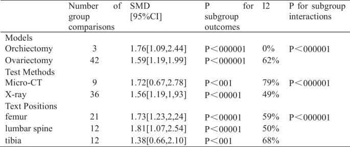 Fig.2 Analysis of BMD in resveratrol subgroups.2
References
- Sun, Jie, et al. "Quercetin attenuates osteoporosis in orchiectomy mice by regulating glucose and lipid metabolism via the GPRC6A/AMPK/mTOR signaling pathway." Frontiers in Endocrinology 13 (2022): 849544. Distributed under Open Access license CC BY 4.0. The image has been modified; only part of the original image. DOI: https://doi.org/10.3389/fendo.2022.849544.
- An, Rongxian, et al. "The effects of resveratrol in animal models of primary osteoporosis: a systematic review and meta-analysis." Journal of Orthopaedic Surgery and Research 19.1 (2024): 137. Distributed under Open Access license CC BY 4.0, without modification. DOI: https://doi.org/10.1186/s13018-024-04595-1.
For Research Use Only.