- Summarize the project requirements and fill in the information collection form.
- Sign a CDA from both parties to further communicate information, such as targets.
- Select an animal model, discuss experimental design, and determine assay parameters.
- Project costing and project schedule forecasting.
Stroke Modeling & Pharmacodynamics Services
Introduction
Stroke, a sudden interruption of blood flow to the brain, stands as a formidable global health challenge, causing significant long-term disability and mortality. This devastating neurological event, primarily categorized as ischemic (due to blockages) or hemorrhagic (due to bleeding), leads to profound neurological deficits and severely impacts quality of life. Understanding its complex pathophysiology and developing effective therapies are paramount.
At Creative Biolabs, we are dedicated to accelerating this critical research by providing a comprehensive suite of well-established and meticulously validated in vivo stroke models, enabling precise evaluation of disease mechanisms and therapeutic interventions.
Available Stroke Models at Creative Biolabs
Animal models of stroke are indispensable for unraveling pathophysiological cascades and assessing neuroprotective and neurorestorative strategies. They enable investigation of brain injury facets like neuronal death, neuroinflammation, and blood-brain barrier disruption in a controlled setting. While no single model fully replicates human stroke heterogeneity, each offers unique insights, crucial for translating research findings into effective clinical treatments.
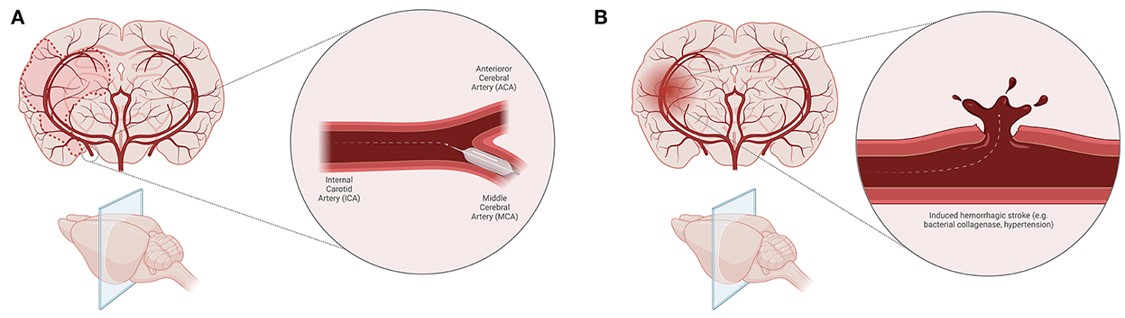 Fig.1 Animal models of ischemic stroke and induced hemorrhagic stroke.1
Our team provides these meticulously designed various stroke models to evaluate disease effects, test therapeutic candidates, and explore underlying mechanisms:
| Stroke Models | Modeling | Animal species |
|---|---|---|
| tMCAO (Transient Middle Cerebral Artery Occlusion) Models | This widely utilized model temporarily occludes the middle cerebral artery via an intraluminal filament. Its transient nature allows reperfusion, mimicking clinical scenarios and proving invaluable for studying reperfusion injury and therapeutic windows of neuroprotective agents. Our standardized protocols ensure high reproducibility. | Mouse, Rat, Dog, NHPs |
| pMCAO (Permanent Middle Cerebral Artery Occlusion) Models | In contrast, permanent MCAO involves sustained middle cerebral artery blockage, often via permanent filament insertion. This model is critical for investigating prolonged cerebral ischemia, evaluating therapies for irreversible brain damage without reperfusion, and providing insights into chronic stroke pathology. | Mouse, Rat, NHPs |
| Photochemically induced Ischemic Stroke Model | This highly controlled focal ischemia model uses an intravenously administered photosensitizing dye and targeted light illumination. This induces localized thrombus formation by damaging endothelial cells. The method offers exceptional precision in controlling lesion size and location, ideal for studying specific cortical areas and microvascular contributions. | Mouse, Rat |
| Collagenase induced Hemorrhagic Stroke Model | Simulating intracerebral hemorrhage (ICH), this model involves stereotaxic injection of bacterial collagenase into brain regions. The enzyme degrades microvessel basal lamina, causing controlled bleeding and hematoma formation. This approach closely mimics human ICH pathology, making it robust for evaluating therapies targeting hemorrhagic stroke injury. | Rat, Dog |
| Sodium Laurate induced Cerebral Microvascular Injury Model | This model induces localized microvascular damage and subsequent ischemic injury. Sodium laurate, a sclerosing agent, is injected into cerebral arteries, causing endothelial injury, thrombosis, and microinfarcts. It provides a valuable tool for investigating microvascular pathology's role in stroke development, particularly relevant for small vessel disease and vascular cognitive impairment. | Rat |
Evaluation Platform
Our preclinical services provide a robust evaluation platform, integrating diverse instruments and tests to characterize stroke outcomes and therapeutic efficacy. We conduct biochemical, molecular, cellular, and histopathological analyses, alongside advanced behavioral assessments and cutting-edge imaging, including but not limited to:
- Infarct volume measurement (magnetic resonance imaging (MRI), tetrazolium chloride (TTC))
- Neurological deficit scores (e.g., Modified Garcia, Bederson)
- Motor and cognitive tests (e.g., rotarod, grip strength, Morris Water Maze)
- Cerebral blood flow and vessels imaging (transcranial laser Doppler)
- Histology analysis (e.g., inflammatory markers)
Applications
Simulating Disease Pathology: Our models accurately simulate the acute and chronic phases of both ischemic stroke (focal and global) and hemorrhagic stroke (intracerebral and subarachnoid hemorrhage), allowing for detailed investigation of lesion progression, neuroinflammation, and long-term functional deficits.
Evaluating Therapeutic Agents: We rigorously evaluate a wide range of therapeutic candidates, including small molecules, biologics, and neuroprotective compounds, assessing their ability to reduce infarct size, improve neurological function, and mitigate secondary brain injury.
Advancing Novel Therapies: Our platforms are ideally suited for assessing innovative neurorestorative strategies, such as stem cell therapy, gene therapy, and rehabilitation interventions, focusing on their potential to promote brain repair, neurogenesis, and functional recovery post-stroke.
Related Cardiovascular Models
Our Advantages
- Diverse Animal Species: While this page focuses on rodent models, we possess expertise across various animal species, allowing for model selection tailored to specific research needs.
- Integrated Evaluation: We offer comprehensive one-stop evaluation services, seamlessly integrating in vivo efficacy studies with in vitro mechanistic investigations.
- Expert Team & Quality Systems: Our professional team comprises highly experienced scientists operating under a perfect management system, ensuring scientific rigor, data integrity, and strict adherence to ethical guidelines.
Work with Us
Inquiry Stage
Project Start
- We provide a detailed project plan, including the required sample quantities, methods and protocols.
- Both parties confirm the project details and start the project.
- Confirm the timeline of the project.
Project Progress
- We provide periodic results and information on the animal's condition.
- We will work together to make project adjustments as necessary.
Project Completion
- We provide a comprehensive project report promptly.
- We arrange transportation for the produced samples.
- We provide a discussion of the project results and help to arrange the next steps.
After-Sales Support
- Data storage and archiving.
Contact Us
With extensive experience in preclinical efficacy studies, Creative Biolabs stands as a reliable research partner, ready to guide your decisions regarding the most suitable stroke model for your unique research objectives. We excel at developing customized study designs and protocols that precisely align with our clients' needs. Please feel free to contact us for more information on the models detailed above or if you wish to propose the development of a novel model.
FAQs
-
Q1: What are the primary differences between ischemic and hemorrhagic stroke models, and why are both important for research?
A: Ischemic stroke models, such as MCAO, simulate blood flow blockage, leading to oxygen and glucose deprivation. Hemorrhagic stroke models, like collagenase-induced ICH, mimic bleeding within the brain. Both are crucial because they represent distinct pathologies with unique injury mechanisms and therapeutic challenges. Studying both types allows for a comprehensive understanding of stroke and the development of targeted treatments for each.
-
Q2: What physiological parameters are typically monitored during acute stroke model induction to ensure model quality and consistency?
A: During acute stroke model induction, we meticulously monitor several critical physiological parameters to ensure model quality and consistency. These commonly include core body temperature, which is maintained within a narrow physiological range to prevent confounding effects on infarct size, as well as blood pressure and blood gas levels to confirm the physiological state of the animal.
-
Q3: Can you customize a stroke model to investigate specific brain regions or a particular stroke subtype?
A: Absolutely. Our scientific team specializes in customizing study designs and protocols. We can modify existing models or develop novel approaches to target specific brain regions, adjust lesion size, or mimic particular stroke subtypes (e.g., cortical vs. subcortical ischemia, specific microvascular injuries) to precisely align with your unique research questions.
-
Q4: What behavioral assessments do you use to evaluate neurological deficits and functional recovery in stroke models?
A: We employ a comprehensive battery of behavioral assessments tailored to the specific model and research goals. For neurological deficits, we frequently use composite scores like the Modified Garcia Score or Bederson Score. For motor function, tests such as the rotarod, grip strength, and locomotor activity are standard. Cognitive recovery is often assessed using tasks like the Morris Water Maze or Novel Object Recognition.
-
Q5: What is the significance of the "therapeutic window" in stroke research, and how do your models help address this?
A: The therapeutic window refers to the limited time frame after stroke onset during which an intervention can be effective. In human ischemic stroke, this window is very narrow (e.g., ~3 hours for permanent focal stroke). Our transient MCAO models, with controlled reperfusion, allow researchers to mimic and investigate interventions within varying therapeutic windows, providing crucial insights into time-sensitive treatments.
-
Q6: Do you offer options for evaluating combination therapies or multi-modal interventions in stroke models?
A: Yes, we are well-versed in designing studies to evaluate complex therapeutic strategies, including combination therapies (e.g., a neuroprotective agent combined with a thrombolytic) or multi-modal interventions (e.g., drug treatment coupled with rehabilitation or cell therapy). Our flexible approach allows for intricate experimental designs to assess synergistic effects.
Published Data
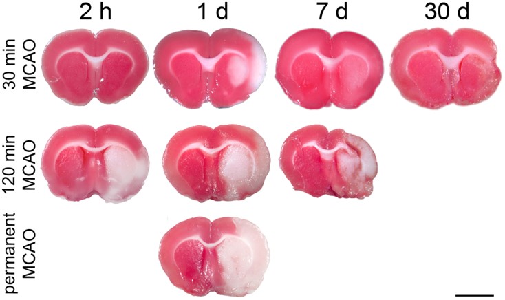 Fig.2 TTC-stained brain sections under the reperfused tMCAO and pMCAO models.2
A compelling example of preclinical stroke research utilizing the MCAO model is demonstrated in the study. This article meticulously characterized ischemic regions in the rats' tMCAO model and pMCAO model. The project results showcased the use of TTC staining to identify infarcts and immunohistochemistry (e.g., for MAP2, HSP72, HSP27) to visualize neuronal damage and stress responses at various time points post-occlusion. This case exemplifies the rigorous methodology employed in MCAO models to precisely assess lesion extent and cellular changes, which is fundamental to evaluating neuroprotective strategies.
References
- Lasica, Nebojsa et al. "Metabolomics as a potential tool for monitoring patients with aneurysmal subarachnoid hemorrhage." Frontiers in neurology vol. 13 1101524. 9 Jan. 2023, DOI:10.3389/fneur.2022.1101524. Distributed under Open Access license CC BY 4.0. The image was modified by extracting and using only part of the original image.
- Popp, Anke et al. "Identification of ischemic regions in a rat model of stroke." PloS one vol. 4,3 (2009): e4764. DOI:10.1371/journal.pone.0004764. Distributed under Open Access license CC BY 4.0, without modification.
For Research Use Only.
Online Inquiry