
Fracture Modeling & Pharmacodynamics Services
Are you currently facing challenges in bone research, such as high variability in healing rates, inconsistent fracture patterns, or the complex interplay between soft tissue trauma and skeletal repair? Our Fracture Modeling & Pharmacodynamics Services help you obtain high-quality, reproducible efficacy data through precision-engineered rat models and multi-dimensional analysis. Creative Biolabs utilizes high-energy impact technologies and stabilizing metatarsal systems to streamline your transition from lead optimization to preclinical validation.
Overview of Fracture Modeling & Pharmacodynamics Services
Fracture modeling involves the creation of standardized skeletal injuries in a controlled laboratory environment to study the physiological stages of bone repair: inflammation, soft callus formation, hard callus mineralization, and remodeling. Bone fractures are a significant global health burden, often complicated by delayed union or non-union due to underlying metabolic conditions, severe trauma, or age-related diseases like osteoporosis. Creative Biolabs' services provide a robust platform to evaluate how pharmacological agents influence these biological pathways, ensuring that potential treatments can effectively restore mechanical and structural integrity to the skeletal system.
Fracture Models
Creative Biolabs specializes in highly reproducible rat models designed to meet various research objectives, ranging from simple bone mineral studies to complex high-energy trauma simulations.
| Models | Related Disease | Drug Evaluation | Animal Species |
| Femoral Fracture Model | Osteoporosis, Osteopenia, Diaphyseal fractures, and age-related bone fragility. | Evaluation of systemic anabolic agents, anti-resorptive drugs (e.g., Bisphosphonates), Sclerostin inhibitors, and hormonal replacement therapies. | Rat |
| Tibial Fracture Model | High-energy open fractures, Non-union/Delayed union, Critical-sized bone defects, and trauma-induced osteonecrosis. | Evaluation of localized growth factors (e.g., BMP-2), angiogenic stimulants, scaffold-based cell therapies (MSCs), and osteoconductive biomaterials. | Rat |
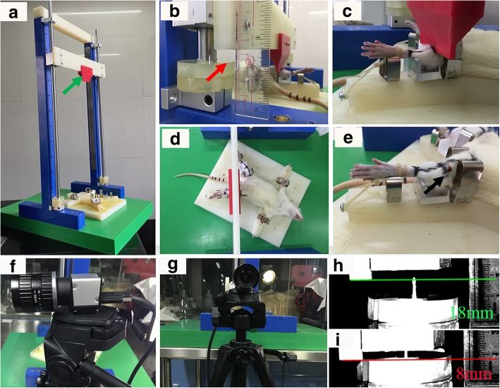 Fig.1 The fracture device and procedure for a representative moderate open fracture (type II).1,3
Evaluation Platform of Our Service
Our platform delivers deep insights through a comprehensive suite of technical parameters, ensuring no biological detail is overlooked during your pharmacodynamics study.
-
Biochemical & Molecular Analysis
- Cytokine Profiling: ELISA-based assays for pro-inflammatory (TNF-α, IL-1β) and anti-inflammatory (TGF-β, IL-10) markers.
- Protein Expression: Western Blot analysis for osteogenic markers (Runx2, Osteocalcin).
- Gene Expression: qPCR for quantifying mRNA levels of bone remodeling factors.
-
Histopathological Examination
- Tissue Staining: H&E and Masson's Trichrome for assessing cellular infiltration and collagen deposition.
- Immunohistochemistry (IHC): Detection of Caspase-3 for apoptosis or CD31 for angiogenesis.
- Scoring Systems: Validated histological grading for fracture union and bone maturity.
-
Behavioral & Functional Testing
- Weight-Bearing Capacity: Dynamic assessment of limb usage using automated pressure plates.
- Gait Analysis: Tracking stride length and paw pressure to evaluate functional recovery.
-
Advanced Imaging & Instrumentation
- Micro-CT (μCT): High-resolution 3D quantification of bone volume (BV/TV) and trabecular microarchitecture.
- DEXA Imaging: Precision Measurement of Bone Mineral Density (BMD) and Mineral Content.
- Biomechanical Testing: Three-point bending and torsion tests to determine the ultimate load and stiffness of the healed bone.
Key Applications
Our models are meticulously designed to simulate specific clinical indications and evaluate a diverse range of therapeutic modalities.
| Indications Simulated: | Evaluated Therapeutic Agents: |
|
|
Why Choose Us?
Creative Biolabs stands as a leader in preclinical orthopedics by providing an unparalleled level of scientific rigor and operational efficiency.
We offer a wide range of validated Rat strains, tailored to your specific metabolic or age-related study requirements.
Our workflow ensures a seamless transition from initial in vitro screening and formulation testing to comprehensive in vivo PD/PK studies, all under one roof.
Our PhD-level team brings decades of experience in orthopedic surgery and molecular biology, supported by rigorous quality management systems that guarantee data integrity for regulatory submissions.
Work with Us
- Summarize the project requirements and fill in the information collection form.
- Sign a CDA from both parties to further communicate information, such as targets.
- Select an animal model, discuss experimental design, and determine assay parameters.
- Project costing and project schedule forecasting.
- We provide a detailed project plan, including the required sample quantities, methods, and protocols.
- Both parties confirm the project details and start the project.
- Confirm the timeline of the project.
- We provide periodic results and information on the animal's condition.
- We will work together to make project adjustments as necessary.
- We provide a comprehensive project report promptly.
- We arrange transportation for the produced samples.
- We provide a discussion of the project results and help to arrange the next steps.
- Data storage and archiving.
Frequently Asked Questions
-
Q: How do you ensure the reproducibility of the fracture pattern in your rat models?
A: At Creative Biolabs, we use precision-engineered apparatus with adjustable buffer discs and high-definition fluoroscopy. This allows us to control the energy transfer and verify the fracture location in real-time, significantly reducing "statistical noise" in your results.
-
Q: Can your platform evaluate systemic effects of a drug, or only local healing?
A: We provide both. Through serum biomarker analysis (ELISA) and multi-organ histopathology, we can assess systemic inflammatory responses alongside the local mechanical recovery of the bone.
-
Q: What is the typical duration for a fracture healing efficacy study?
A: Most studies run between 3 to 7 weeks. We perform weekly weight-bearing tests and longitudinal imaging to provide you with a chronological map of the drug's efficacy. Reach out to us for a customized timeline for your specific molecule.
-
Q: How do you handle animal welfare and pain management in these trauma models?
A: Creative Biolabs adheres to the highest ethical standards. All procedures include systematic analgesia and post-operative care protocols that ensure animal well-being without interfering with the inflammatory phase of bone healing.
Published Data
Objective: To assess the efficacy of high-bioavailability Amorphous Calcium Carbonate (ACC) on femoral fracture healing compared to standard crystalline calcium.
Model Used: Male Wistar Rat mid-femur fracture model with internal needle stabilization.
Results: The intramedullary-stabilized femoral fracture model demonstrated exceptional sensitivity and reproducibility for quantifying the pro-regenerative effects of ACC. Results exhibited a precise dose-dependent escalation in biomechanical rigidity and functional weight-bearing symmetry. High-resolution Micro-CT revealed accelerated callus mineralization, while biochemical profiling confirmed a significant reduction in bone resorption markers within 14 days. By effectively mimicking clinical stabilization, this robust model provides a high-fidelity translational platform, offering multi-dimensional readouts, from micro-architectural preservation to functional recovery, essential for validating novel systemic osteogenic therapeutics.
Creative Biolabs provides the expertise and advanced instrumentation necessary to validate your bone-healing therapeutics with confidence. From high-energy tibial trauma to standardized femoral modeling, we deliver data that drives decisions. Contact our team; we are available to provide detailed protocols and pricing for your specific research needs.
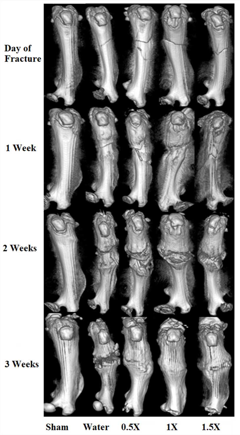 Fig.2 Micro-CT visualizations of femoral bones in study cohorts at 1,2 and 3weeks post-fracture.2,3
References
- Shi, Enxian, et al. "A novel rat model of tibial fracture for trauma researches: a combination of different types of fractures and soft tissue injuries." Journal of Orthopaedic Surgery and Research 14.1 (2019): 333. DOI: https://doi.org/10.1186/s13018-019-1386-4.
- Yeh, Tsu-Te, et al. "Amorphous Calcium Carbonate Enhances Fracture Healing in a Rat Fracture Model." Nutrients 16.23 (2024): 4089. DOI: https://doi.org/10.3390/nu16234089.
- Distributed under Open Access license CC BY 4.0, without modification.
For Research Use Only.