Retinal Fibrosis Modeling & Pharmacodynamics Services
Introduction
Retinal fibrosis develops into scar tissue formation which progressively diminishes the retinal function. The disease starts with subtle symptoms but progresses to cause blurred vision alongside light sensitivity while creating floaters and narrowing visual fields. Elevated intraocular pressure and retinal detachment trigger retinal fibrosis development whereas diseases such as diabetic retinopathy and rare ocular tumors also initiate this condition. Researchers often employ rodents such as mice, rats, and rabbits as their animal models in scientific studies. Animal disease models serve as representations of human disease pathways and progression which help researchers gain insights into disease origins as well as mechanisms and pathophysiology. Creative Biolabs employs animal models to recreate human retinal fibrosis development and its risk factors and pathogenesis to advance drug development for treating this disease and enhance treatment approaches.
Retinal Fibrosis Disease Models Available at Creative Biolabs
- Two-stage Laser-induced Subretinal Fibrosis Secondary to CNV
Subretinal fibrosis acts as the healing response following choroidal neovascularization development in neovascular age-related macular degeneration. The development of subretinal fibrosis results in the destruction of local photoreceptors along with retinal pigment epithelium and choroidal vasculature which causes permanent macular visual system malfunction. The experimental study involves two stages with C57BL/6J mice who are between 2 to 3 months old. The initial step involves performing laser surgery to treat CNV. A second laser burn targeting CNV lesions formed one week earlier is applied seven days following the first stage (Day 0). The fundus examination reveals a visible white/yellow spot. The laser settings matched those applied during the initial laser treatment. Fibrotic lesions develop over 40 days. Researchers employ fundus and optical coherence tomography (OCT) imaging methods to non-invasively monitor lesion development. Researchers use Image J software to measure lesion numbers and areas whereas retinal tissue sections and Western blots provide detailed lesion descriptions.
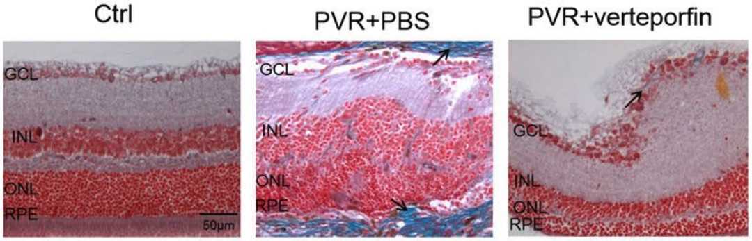 Fig. 1 Representative images of retinas of PVR mouse models stained by Masson.1
- Proliferative Vitreoretinopathy (PVR) Models
Proliferative vitreoretinopathy (PVR) emerges as a serious complication from rhegmatogenous retinal detachment (RRD), which leads to vision loss. Retinal tears from RRD-induced blood-retinal barrier disruption affect retinal pigment epithelial (RPE) cells located between the neurosensory retina and Bruch's membrane via multiple cytokines including VEGF, TGF-β, and PDGF-BB. Following retinal detachment exposure to cytokines RPE cells activate and transition through epithelial-to-mesenchymal transition (EMT). Through the process of EMT RPE cells transform into myofibroblasts that create contractile subretinal and epiretinal membranes. The contraction of these membranes triggers tractional retinal detachment. Researchers choose animal species for PVR models by evaluating both the appropriateness of the animal and how manageable the experiments are. Scientists often select rabbits for these experiments because their small lenses enable intraocular procedures to be performed without damaging the lens or retina. The research community has extensively used rabbit models to perform intravitreal injections of fibroblasts or retinal pigment epithelial (RPE) cells. Mice present certain challenges for PVR models due to their large lenses and small vitreous volume which complicate surgical manipulation, yet they remain valuable for research because of the multiple available transgenic mouse models. Transgenic mice models supply abundant genetic data which supports more detailed investigations into PVR pathogenesis.
Measurements
Creative Biolabs provides retinal fibrosis model detection services, covering various testing and analysis methods, including:
- Lens opacity
- Intraocular pressure
- Optical coherence tomography
- Western blotting analysis (e.g. Tau, NOX1, NOX2, NOX4, NOXO1, p47PHOX)
- Histopathological observation (e.g. retina, optic nerve, cornea)
Related Ocular Disease Models
Moreover, you can find various other examples of rodent ocular disease models, including a range of conditions used for testing and research purposes, such as:
- Dry Eye Models
- Corneal Disease Models
- Cataract Models
- Glaucoma Models
- Dry Age-Related Macular Degeneration (AMD) Models
- Wet Age-Related Macular Degeneration (AMD) Models
- Fundus Disease Models
- Diabetic Retinopathy Models
- Retinal Vein Occlusion Models
- Ocular Inflammation Models
Creative Biolabs has developed a thorough evaluation platform for disease animal models. For inquiries, please refer to the contact details provided on the company's homepage.
Reference
- Zhang, Wei, and Jing Li. "Yes‐associated protein is essential for proliferative vitreoretinopathy development via the epithelial‐mesenchymal transition in retinal pigment epithelial fibrosis." Journal of Cellular and Molecular Medicine 25.21 (2021): 10213-10223. Distributed under Open Access license CC BY 4.0. The image was modified by extracting and using A part of the original image.
For Research Use Only.