Diabetic Retinopathy Modeling & Pharmacodynamics Services
Introduction
Diabetic retinopathy (DR) stands for the specific eye disease that develops in individuals with diabetes mellitus (DM). Diabetic retinopathy development involves vascular and neuronal systems alongside inflammatory processes that activate complex cellular and molecular signaling pathways. Untreated this disease can cause vision loss followed by complete blindness. Researchers have created animal models that replicate various diabetic complications to support their investigations. The animal models for research purposes consist of pharmacologically induced hyperglycemia models, natural diabetic rodent models, and vascular growth models without diabetes. Creative Biolabs utilizes animal models to understand DR development and progression along with its risk factors and pathogenesis while aiding in drug discovery and therapeutic optimization.
Diabetic Retinopathy Disease Models Available at Creative Biolabs
- Streptozotocin-Induced Diabetic Retinopathy Models
Diabetic retinopathy begins when high blood sugar levels generate microvascular damage and enhance vascular permeability which then leads to diabetic macular edema and capillary blockage. Retinal capillary blockage results in retinal ischemia which then elevates VEGF levels leading to neovascularization. STZ administration stands as the preferred method for creating diabetic retinopathy models because this approach produces the quickest progression of the disease. C57Bl/6 mice showed a rise in novel glial cell numbers and a decrease in retinal ganglion cells following intraperitoneal STZ administration through both single high doses and multiple low doses.
- Streptozotocin-Induced Diabetic Cataract Models
About 20% of diabetic patients develop cataracts which represent one of the condition's most frequent complications. The longer diabetes alongside elevated blood glucose levels elevates the risk of developing and worsening cataracts. Diabetic cataracts may involve three mechanisms: The development of diabetic cataracts occurs due to increased polyol pathway activity combined with non-enzymatic lens protein glycation and oxidative stress conditions. Diabetic complications arise from oxidative stress which acts as a key element in diabetic cataract formation. The administration of STZ through intravenous injection caused rats to display typical diabetic symptoms rapidly. Lens changes to posterior striped opacity were seen in the 4th week and by the 16th week, all rats developed progressive cataract formation which led to severe cataracts and bilateral blindness. The retina displayed distinct circular blood vessels paired with microaneurysm formation.
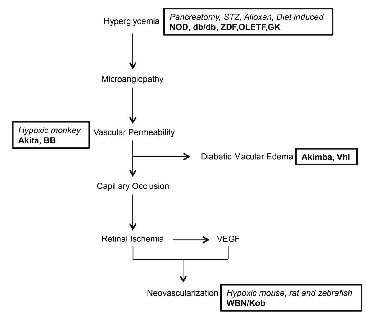 Fig. 1 Schematic representation of diabetic retinopathy (DR) disease progression.1,3
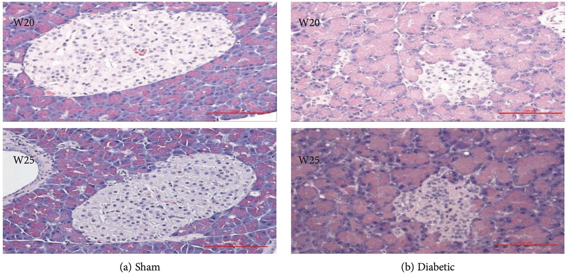 Fig. 2 Representative images of the pancreas from rats 20 and 25 weeks after STZ or sham injection stained with HE.2,3
- Diabetic Macular Edema Models
Diabetic macular edema represents a common complication occurring in diabetes patients. Visual acuity becomes blurred when the damaged blood vessels leak fluid because impaired vascular endothelial barrier function leads to edema. The streptozotocin (STZ)-induced DME model demonstrates clinical retinal alterations including increased retinal thickness and macular edema. The quantification of multiple pro-angiogenic and pro-inflammatory markers enables researchers to objectively evaluate how successful a drug is in stopping pathological blood vessel growth while also reducing inflammation. The variations in these biomarkers function as therapeutic efficacy indicators that assist in determining treatment effectiveness and safety for diabetic macular edema.
Measurements
With the well-established diabetic retinopathy model, Creative Biolabs can provide customers with drug candidate efficacy evaluation services, drug mechanism exploration, and verification. The test parameters are listed below:
- Transmission electron microscopy (TEM)
- Optical Coherence Tomography (OCT)
- Immunohistochemistry (IL-6, TNF-α, VEGF)
- Spectral-domain optical coherence tomography (SD-OCT)
- Fluorescein angiography (FA)
Related Ocular Disease Models
Except for the Diabetic Retinopathy models, we can also provide the following other ocular disease models to our global customers.
- Dry Eye Models
- Corneal Disease Models
- Cataract Models
- Glaucoma Models
- Dry Age-Related Macular Degeneration (AMD) Models
- Wet Age-Related Macular Degeneration (AMD) Models
- Fundus Disease Models
- Retinal Fibrosis Models
- Retinal Vein Occlusion Models
- Ocular Inflammation Models
Thanks to many outstanding scientists and perfect experimental platforms, Creative Biolabs has established a variety of animal models, providing high-quality efficacy evaluation services to a large number of customers. You can contact us at any time to discuss your project and design.
References
- Olivares, Ana Maria, et al. "Animal models of diabetic retinopathy." Current diabetes reports 17 (2017): 1-17.
- Wang-Fischer, Yanlin, and Tina Garyantes. "Improving the reliability and utility of streptozotocin‐induced rat diabetic model." Journal of diabetes research 2018.1 (2018): 8054073.
- Distributed under Open Access license CC BY 4.0, without modification.
For Research Use Only.