Corneal Disease Modeling & Pharmacodynamics Services
Introduction
Corneal injury is mainly caused by diabetic keratopathy, inflammation after corneal chemical burns, and corneal endothelial damage caused by trauma. It is difficult to treat and seriously affects the patient's vision. The cornea is divided into five layers, including the epithelial layer, the posterior elastic layer, which can be regenerated after injury, and the anterior elastic layer, the stromal layer, and the endodermis, which cannot be regenerated after injury. The animal model is an important means to study the regulatory mechanism and explore and evaluate its prevention and treatment methods. Creative Biolabs has developed a range of proven corneal disease models to help our customers more effectively explore the mechanisms of corneal disease and develop research programs for efficacy.
Corneal Disease Models Available at Creative Biolabs
Corneal Neovascularization Models
Corneal neovascularization (CNV) frequently results in the loss of normal corneal transparency, a significant contributor to visual impairment and a major risk factor for post-corneal transplantation rejection. CNV animal model is an animal model of induced disease, commonly used in rats and rabbits. It induces neovascularization in the animal cornea through physical, chemical, biological, and surgical means, producing a condition similar to human diseases.
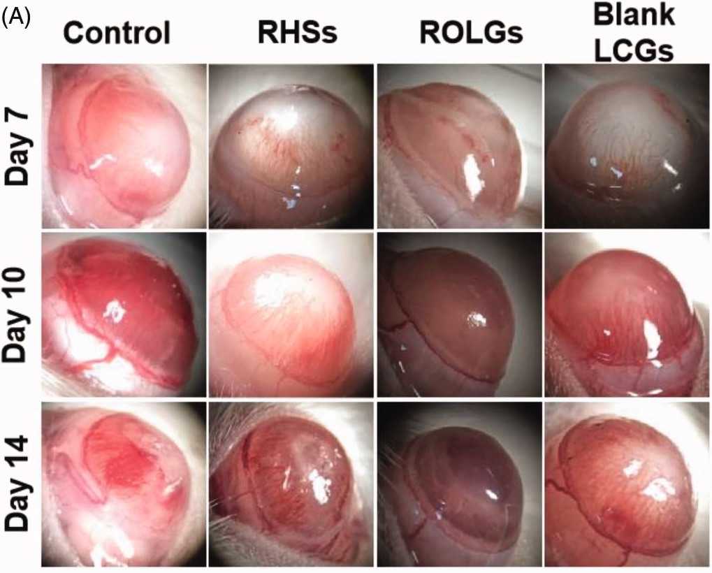 Fig. 1 Images of alkaline injury rat cornea model.1
- Corneal suture method
This method is simple and easy to operate, which is beneficial for observing and comparing the growth of neovascularization. It can exclude the influence of chemical drugs on the prevention and treatment of drugs. It has little damage to the cornea, and it is easier to prevent and treat postoperative infection than other methods. It can better simulate the pathological development process of corneal neovascularization formed by clinical inflammatory stimulation.
- Alkali burn method
The alkali burn method is a traditional method for establishing CNV, which belongs to the inflammatory model. After burning the animal cornea with NaOH filter paper, neutrophils infiltrate the corneal stroma, and the synthesis of local arachidonic acid derivatives increases, resulting in increased vascular permeability, vasodilation, and induction of neovascularization.
Corneal Wound Healing Models
The corneal injury model is established by acupuncture injury of the rat cornea. This model can better control the degree of corneal injury and is easy to operate. In addition, the acupuncture injury model not only damages the corneal epithelium but also damages the corneal basement membrane and anterior stromal layer.
Measurements
Creative Biolabs can provide detection services for corneal disease models, such as:
- Immunohistochemical detection (e.g., BMP-2, BMP-7)
- Observation by slit lamp microscope (The growth of CNV, corneal opacity score, diameter of corneal epithelial defect)
- Histopathological observation (e.g., cornea, conjunctiva, lacrimal gland)
Related Ocular Disease Models
Additionally, we also offer other ocular disease models, such as:
- Dry Eye Models
- Cataract Models
- Glaucoma Models
- Dry Age-Related Macular Degeneration (AMD) Models
- Wet Age-Related Macular Degeneration (AMD) Models
- Fundus Disease Models
- Diabetic Retinopathy Models
- Retinal Fibrosis Models
- Retinal Vein Occlusion Models
- Ocular Inflammation Models
Creative Biolabs has a comprehensive preclinical efficacy evaluation platform to help new drugs' clinical transformation. Our contact information can be located on the company's homepage.
Reference
- Li, Minshu, et al. "Ocular lamellar crystalline gels for sustained release and enhanced permeation of resveratrol against corneal neovascularization." Drug Delivery 28.1 (2021): 206-217. Distributed under Open Access license CC BY 4.0. The image was modified by extracting and using A parts of the original image.
For Research Use Only.