Wet Age related Macular Degeneration (AMD) Modeling & Pharmacodynamics Services
Introduction
Age-related macular degeneration (AMD) progressively destroys the macular area and represents a leading cause of visual disability among people aged 50 and older across the globe. AMD mainly has two types: AMD presents two distinct forms which are the neovascular type called "wet" AMD and the non-neovascular type named "dry" AMD. Bruch's membrane disruption due to wet AMD leads to macular neovascularization (MNV) and results in retinal hard exudates together with subretinal hemorrhage and serous retinal detachment and scarring. Animal models provide essential research tools for wet AMD studies that fuel drug development advancements. Creative Biolabs uses animal models to study AMD development alongside its risk factors and pathogenic mechanisms. Through our advanced ophthalmology platforms, we deliver extensive preclinical drug assessment services to global clients which aid both drug development and disease treatment improvements.
Dry AMD Disease Models Available at Creative Biolabs
Laser-Induced Choroidal Neovascularization (CNV) Models
The mouse laser-induced choroidal neovascularization model serves as an essential foundation for wet AMD research. The retinal pigment epithelium (RPE) layer along with Bruch's membrane sustains significant damage at the photocoagulation site during the initial stages following laser treatment in mice. The formation of large lumen neovascularization occurs underneath the retina one-week following photocoagulation. Abnormal proliferation of RPE cells occurs while numerous inflammatory cells infiltrate the area surrounding CNV.
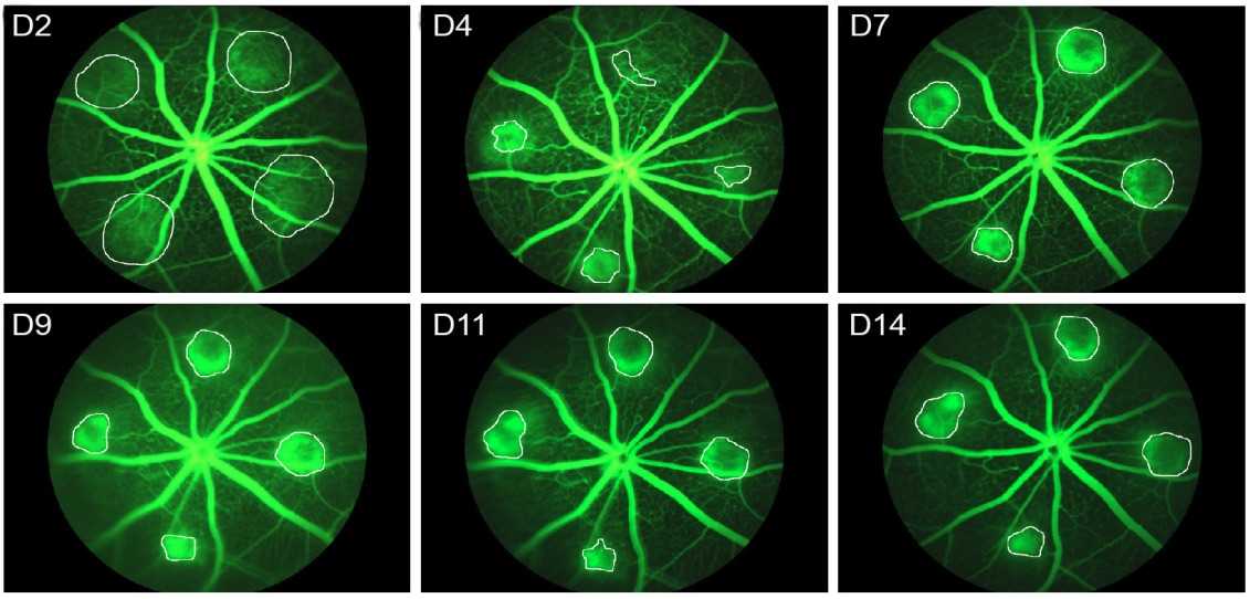 Fig. 1 Representative fluorescent images of CNV mouse model.1
DL-alpha-aminoadipic acid (DL-AAA)-Induced Retinal Neovascularization and Leakage Models
The DL-AAA is a retinal glial cell toxin that has been demonstrated to induce retinal neovascularization and chronic leakage in the retinal vasculature. The symptoms of this model are very similar to those of patients with advanced wet AMD. After a single intravitreal (IVT) injection of DL-AAA in rabbits, retinal neurons degenerated, and retinal neovascularization (RNV) formed in injury. This morphology and leakage remained stable at 10 weeks and persisted for at least 65 weeks.
VEGF-Induced Retinal Leakage Models
Vascular endothelial growth factor (VEGF) functions as an extremely potent protein for increasing vascular permeability. Wet AMD models show that when VEGF is injected into the vitreous cavity it causes time- and dose-dependent breakdown of both the blood-retinal and blood-aqueous barriers. Multiple retinal effects occur due to VEGF including substantial vasodilation as well as vascular tortuosity along with fluorescein leakage from retinal vessels and intraretinal edema formation. The primary treatment approach for wet AMD involves anti-VEGF therapy.
Other Models
AMD pathogenesis includes oxidative stress together with immune inflammation as well as the metabolism of both lipids and carbohydrates. Screening identified multiple related candidate genes which allowed scientists to establish AMD mouse models. Subretinal neovascularization and hyperreflective lesions resembling subretinal fibrosis in human patients emerge in JR5558 transgenic mice. The HUVEC tube formation assay represents a straightforward yet established method for studying angiogenesis in vitro through endothelial cell formation of tubular structures like capillaries on growth factor-reduced basement membrane extract gel. They serve as models for Wet AMD to test potential pharmaceutical compounds.
Measurements
Creative Biolabs can provide testing parameters for wet age-related macular degeneration models, such as:
- Color fundus photography
- Transmission electron microscopy (TEM)
- Optical Coherence Tomography (OCT)
- Immunohistochemistry (GFAP, VEGF, RPE-65, Collagen IV)
- Spectral-domain optical coherence tomography (SD-OCT)
- Fluorescein angiography (FA)
- Scanning laser ophthalmoscopy (SLO)
Related Ocular Disease Models
Except for the wet AMD models, we can also provide the following other ocular disease models to our global customers.
- Dry Eye Models
- Corneal Disease Models
- Cataract Models
- Glaucoma Models
- Dry Age-Related Macular Degeneration (AMD) Models
- Fundus Disease Models
- Diabetic Retinopathy Models
- Retinal Fibrosis Models
- Retinal Vein Occlusion Models
- Ocular Inflammation Models
Creative Biolabs has established a comprehensive preclinical drug efficacy evaluation platform. You can contact us at any time if you have any needs.
Reference
- Salas, Anna, et al. "Neovascular progression and retinal dysfunction in the laser-induced choroidal neovascularization mouse model." Biomedicines 11.9 (2023): 2445. Distributed under Open Access license CC BY 4.0, without modification.
For Research Use Only.