
Fluorophore Conjugated ASO
Fluorophore-conjugated ASOs enable direct visualization and quantitative tracking of oligonucleotide uptake, intracellular distribution, trafficking, and tissue accumulation. Reliable results depend on selecting a fluorophore, linker, and conjugation site that preserve ASO hybridization, nuclease resistance, cellular uptake, and biological activity while minimizing signal interference and dye-related artifacts. Each conjugate should therefore be validated for labeling efficiency, purity, fluorescence stability, target engagement, and functional performance.
Introduction
Fluorophore-conjugated ASOs are labeled antisense oligonucleotides that combine sequence-specific RNA targeting with fluorescent detection. They are used to visualize and measure ASO uptake, distribution, intracellular trafficking, localization, and degradation in cells and tissues. Creative Biolabs provides a comprehensive fluorophore-conjugated ASO development service, covering labeling design, conjugation, purification, cellular tracking, and in vivo distribution analysis.
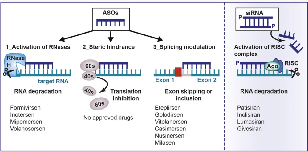 Figure 1. Mechanism of action of antisense oligonucleotides.1
A Fluorophore Is Both Reporter and Chemical Modification
The reporter can change the molecule it is intended to observe
Common organic dyes can add hydrophobicity, negative or positive charge, and a relatively bulky aromatic system. Those properties may alter reverse-phase retention, plasma-protein association, membrane binding, endosomal localization, renal handling, or aggregation. A label at one terminus may be tolerated while the same dye at an internal site interferes with hybridization or protein recruitment.
Fluorescence depends on environment and molecular state
Guanine-proximal dyes may be quenched, crowded dyes may self-quench, and acidic vesicles can suppress pH-sensitive reporters. Tissue scattering, absorption, and autofluorescence further distort in vivo signals. A bright pixel can represent intact ASO, a short fluorescent metabolite, free dye, or locally dequenched material. Conversely, low signal can reflect quenching or photobleaching rather than absence of ASO.
| Observed Signal | Possible Molecular State | Alternative Explanation | Resolving Measurement |
|---|---|---|---|
| Bright puncta in cells | Endosomal conjugate or concentrated fragments | Surface-bound probe or dye aggregation | Surface stripping, z-stack imaging, organelle markers, intact-species assay |
| Diffuse cytosolic signal | Released or dispersed ASO-containing species | Free dye or photoproduct | Metabolite separation and unlabeled functional readout |
| Low fluorescence after formulation | Quenched carrier-associated probe | Poor loading or chemical loss | Detergent/chaotrope recovery and mass balance |
| Persistent tissue signal | Retained intact conjugate | Stable fluorescent metabolite or autofluorescence | Chromatographic identity plus blank-tissue correction |
Choosing a Dye Around the Measurement
- For flow cytometry, prioritize detector compatibility, compensation against other fluorophores, and a signal range that does not saturate at early time points.
- For confocal or super-resolution microscopy, balance photon output, photobleaching, localization precision, and compatibility with fixation or live-cell conditions.
- For whole-animal imaging, choose far-red or near-infrared excitation and include tissue-specific calibration because depth and optical properties differ among organs.
- For hybridization-sensitive designs, evaluate sequence-dependent quenching and fluorescence changes in single-stranded, duplex, protein-bound, and degraded states.
- For quantitative comparisons, keep dye, labeling position, degree of labeling, acquisition settings, and sample processing identical across candidates.
| Research Question | Useful Optical Property | Primary Confounder | Minimum Control |
|---|---|---|---|
| How much probe associates with cells? | Bright, stable visible or far-red label | Surface binding counted as uptake | Temperature or stripping control plus internalization assay |
| Where does the probe traffic? | Photostable dye compatible with organelle markers | Spectral bleed-through and fixation redistribution | Single-color controls and live/fixed comparison |
| Where does material distribute in vivo? | Near-infrared emission and low autofluorescence | Depth, scattering, and fluorescent metabolites | Ex vivo calibration and intact-species quantification |
| Does hybridization occur? | Environment- or proximity-sensitive reporter pair | Sequence quenching unrelated to target binding | Mismatch target and dye-position controls |
Label Placement and Conjugation Strategy
Terminal labels simplify product definition
A 5-prime or 3-prime label usually provides a defined one-to-one construct and minimizes disruption of internal base pairing. The preferred terminus depends on the ASO mechanism and required end functionality. A terminal dye can still affect exonuclease processing, protein interactions, or uptake, and cleavage can separate it from most of the ASO. A hydrophilic spacer may reduce steric interference and dye-base stacking, but longer spacers add flexibility and possible heterogeneity. The parent ASO and a spacer-only intermediate help identify whether observed changes originate from the dye or attachment architecture.
Internal and base-analogue labels answer specialized questions
Internal amino-modified nucleotides support site-specific labeling, dual-label geometries, or probes that retain a required terminal group. Placement within the recognition sequence can alter duplex stability and RNase H compatibility, so position-matched activity testing is essential. Fluorescent base analogues offer a smaller, in-sequence alternative and can preserve gapmer behavior in selected contexts, but their brightness is often lower and multiple incorporations may perturb structure or activity. Dual labels add FRET, quenching, or cleavage readouts while increasing synthesis, purification, spectral correction, and interpretation demands.
The coupling route must fit dye and ASO stability
Direct phosphoramidite incorporation can give a defined label during solid-phase synthesis when the dye tolerates coupling, oxidation, and deprotection. Post-synthetic amine-NHS ester, thiol-selective, or click reactions accommodate sensitive reporters and modular dye panels. Each route can leave unreacted ASO, free dye, hydrolyzed reagent, positional isomers, or incompletely labeled material. Custom oligonucleotide modification can introduce controlled handles, but the final conjugate still requires purification and orthogonal identity confirmation rather than reliance on absorbance ratios alone.
From Photons to Biological Meaning
Interpretation is strongest when the experiment follows a chain from administered material to RNA response. Skipping a link encourages fluorescence to be mistaken for productive delivery.
- Confirm the administered article: sequence, dye, attachment position, purity, free dye, aggregation, and fluorescence response in the dosing matrix.
- Measure extracellular state: stability, protein or carrier association, quenching, and recovery after dilution or disruption.
- Separate cell-surface binding from internalization using temperature shifts, competition, stripping, or microscopy with optical sectioning.
- Resolve subcellular location with validated organelle markers, spectral controls, time courses, and methods that distinguish punctate from diffuse signal.
- Determine whether fluorescence remains linked to intact ASO through chromatography, mass spectrometry, hybridization capture, or size-resolved assays.
- Measure target RNA and a mechanism-matched protein, splice, or phenotypic endpoint with unlabeled and labeled constructs at matched ASO molar dose.
Experimental Designs for Different Questions
Cell association and trafficking require different readouts
Flow cytometry efficiently compares fluorescence per cell but generally cannot distinguish membrane-bound, endosomal, and released species without additional procedures. Imaging adds spatial information, yet colocalization coefficients depend on resolution, thresholds, marker choice, and time. Live-cell imaging avoids some fixation artifacts but increases phototoxicity and bleaching. A robust uptake study combines an unlabeled biological endpoint with at least two orthogonal fluorescent measurements. ASO in vitro screening can be structured around free uptake rather than transfection, since transfection reagents bypass the delivery barrier that the label is meant to reveal.
In vivo fluorescence is semi-quantitative until calibrated
Whole-animal imaging is useful for time-resolved localization and candidate triage, but depth, fur, hemoglobin, tissue scattering, and detector geometry prevent simple concentration comparisons across organs. Ex vivo organ imaging reduces some variation without identifying intact ASO or cell type. Tissue homogenization with matrix-matched standards, recovery procedures that reverse quenching, and an orthogonal chemical assay improve quantification. Cell sorting or spatial imaging can then test whether signal reaches the intended population. In vivo antisense studies should connect fluorescence with intact exposure, target modulation, and tolerability.
- Use instrument settings below saturation and retain raw acquisition parameters for all groups and time points.
- Include blank tissue, unlabeled ASO, free dye, labeled parent, sequence control, and delivery-vehicle controls as appropriate.
- Report fluorescence per cell or tissue together with viability, recovery, and normalization method rather than as arbitrary units alone.
- Use washout and pulse-chase designs to separate rapid association from retention, processing, and delayed RNA activity.
Quality Controls
The most informative control set varies with the claim. A labeled sequence control tests sequence-dependent localization but not dye perturbation. An unlabeled parent tests activity but is invisible. Free dye tests nonspecific fluorescence but may have distribution unlike a cleaved dye-bearing fragment. A spacer-only ASO, dye-position variant, noncleavable linker, and deliberately cleaved standard address other hypotheses. For receptor or carrier studies, competition and component-only controls establish mechanism. No single control can resolve chemical identity, optical behavior, uptake route, and pharmacology simultaneously.
| Claim | Essential Comparison | Orthogonal Evidence | Decision Criterion |
|---|---|---|---|
| Label preserves ASO function | Labeled versus unlabeled parent at matched molarity | Target RNA and protein/splice endpoint | Comparable concentration-response and mechanism |
| Signal represents intact probe | Intact, cleaved, and free-dye standards | Chromatography or hybridization capture | Major fluorescence co-resolves with intact species |
| Cellular uptake is productive | Sequence control and uptake-pathway perturbation | Subcellular localization plus RNA response | Internalization precedes sequence-dependent activity |
| Tissue fluorescence reflects exposure | Matrix-matched organ standards and blank tissue | Chemical ASO quantification and cell resolution | Calibrated signal tracks intact target-cell material |
| Two candidates can be compared | Same dye, site, dose, processing, and settings | Recovery and photophysics in each matrix | Difference exceeds technical and matrix variability |
Characterization and Stability of the Labeled ASO
Chemical identity and optical identity are separate attributes
Label incorporation must be confirmed by mass spec, while chromatography quantifies full-length ASO, unlabeled parent, and fragments. UV-Vis and fluorescence spectra reveal label behavior but require complementary methods; a single purity percentage is insufficient.
Stability studies must track linkage and signal behavior
Stability studies must track chemical composition and fluorescence output under stress (light, freeze-thaw, nucleases, etc.) using methods that quantify intact conjugate, fragments, free dye, and photobleaching. Container adsorption and recovery controls in biological matrices are essential to avoid misinterpreting signal loss.
Limits of Fluorescence-Based ASO Tracking
Fluorescence tracks localization and kinetics but does not prove antisense mechanism, chemical integrity, molecular interaction, target-cell exposure, or durable activity. Dyes can alter biodistribution, so conclusions must be bounded by matched pharmacology, analytics, matrix calibration, cell-resolution, and RNA endpoint data.
Frequently Asked Questions
Q: Does a fluorescent label always preserve ASO behavior?
A: No. A dye can change hydrophobicity, charge, protein binding, uptake, stability, or RNA activity. Compare the labeled construct with the identical unlabeled parent.
Q: Which labeling position is usually least disruptive?
A: A terminal 5-prime or 3-prime position is often easier to characterize than an internal site, but the preferred terminus depends on ASO mechanism, end chemistry, linker, and assay.
Q: Can fluorescence quantify intact ASO in tissue?
A: Not by itself. Fluorescence can arise from intact conjugate, labeled fragments, free dye, or dequenched material. Matrix calibration and an orthogonal intact-species assay are needed.
Q: How should a fluorophore be selected for in vivo imaging?
A: Match excitation and emission to the instrument and tissue optical window, then evaluate brightness, charge, photostability, pH response, protein effects, and matrix-specific recovery.
Q: What controls distinguish surface binding from internalization?
A: Use temperature or uptake-pathway perturbation, extracellular fluorescence quenching or surface stripping, optical sectioning, washout time courses, and validated subcellular markers.
Q: Are fluorescent base analogues better than external dyes?
A: They can be less perturbing in selected ASO designs, but they are often dimmer and can still affect duplex structure or activity. Suitability must be tested for the specific sequence and assay.
Published Data
Case 1: Magnetic-Guided Delivery of Cy3-Labeled ASOs in Retinal Explants and Organoids
This study utilizes Cy3 fluorophore-labeled antisense oligonucleotides (ASO-Cy3) to validate magnetic nanoparticle (MNP)-guided delivery across human retinal organoid (hRO) and explant models. To optimize ocular ASO delivery, researchers combined MNP carriers with external magnetic guidance to enhance targeted transduction efficiency. Using ASO-Cy3 enabled direct tracking and precise quantification of tissue penetration and intracellular uptake via confocal 3D Z-stack imaging, flow cytometry, and immunofluorescence—co-localizing red Cy3 signals with green CRX-positive photoreceptor precursor markers. Quantitative analyses confirmed that magnetic guidance significantly accelerated uptake kinetics, heightened Cy3 fluorescence intensity, and increased the percentage of Cy3-positive retinal cells compared to unguided controls. This study illustrates a practical paradigm for fluorophore-conjugated ASOs, demonstrating how fluorescent labeling serves as an indispensable analytical tool for visual tracking, quantitative spatial mapping, and optimizing targeted nucleic acid delivery in complex three-dimensional organoid platforms.
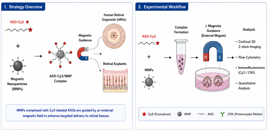 Figure 2. Cy3-ASO delivery via magnetic guidance in retinal tissues and organoids.
Overview of What Creative Biolabs Can Provide
Creative Biolabs can support fluorophore-ASO studies from parent sequence preparation and labeling-site selection through conjugation, purification, optical characterization, cellular tracking, and in vivo distribution analysis. The modules below correspond to the probe-design and interpretation questions addressed in this resource.
| Research Need | Related Creative Biolabs Support | How It Connects to the Current Resource Topic |
|---|---|---|
| Develop a purpose-built labeled ASO | Fluorophore-Conjugated Antisense Oligonucleotide (ASO) Development Service | Integrates dye selection, attachment position, linker, purification, optical verification, and functional comparison. |
| Compare alternative ASO conjugation formats | Antisense Oligonucleotide (ASO) Conjugate Development Services | Places fluorophore labeling alongside delivery-oriented peptide, aptamer, antibody, lipid, and other conjugates. |
| Select a compatible reporter chemistry | Fluorophores | Provides dye options that can be matched to spectral range, instrumentation, matrix, and labeling handle. |
| Install a defined labeling handle | Custom Oligonucleotide Modification Service | Introduces terminal or internal functionality for site-specific post-synthetic conjugation. |
| Prepare matched parent and control ASOs | Custom Antisense Oligonucleotide Synthesis | Supplies unlabeled, sequence-control, spacer-control, and label-ready oligonucleotides for attribution. |
| Evaluate uptake and RNA activity in vitro | Antisense Oligonucleotide (ASO) In Vitro Screening Service | Combines cellular fluorescence with free-uptake, viability, target RNA, and mechanism-matched functional readouts. |
| Assess distribution and pharmacology in vivo | In Vivo Study Service for Antisense Therapeutics | Connects calibrated imaging with intact exposure, cell-resolved distribution, target modulation, and tolerability. |
To discuss dye selection, labeling position, calibration, or biological tracking for your ASO, contact us today to connect with our scientific team.
References
- Collotta D, Bertocchi I, Chiapello E, et al. Antisense oligonucleotides: a novel Frontier in pharmacological strategy. Frontiers in pharmacology, 2023, 14: 1304342. https://doi.org/10.3389/fphar.2023.1304342 Distributed under Open Access license CC BY 4.0, with modification.
- Ye X, Chen S, Xiong W, et al. Magnetic-Guided Delivery of Antisense Oligonucleotides for Targeted Transduction in Multiple Retinal Explant and Organoid Models. Advanced Science, 2025, 12(22): 2417363. https://doi.org/10.1002/advs.202417363
