
Lipid Conjugated ASO
Lipid-conjugated ASOs can improve tissue exposure, membrane interaction, cellular uptake, and intracellular release while enabling tunable, chemically defined delivery without the complexity of nanoparticles. They are particularly useful for targeted gene knockdown when lipid–protein or lipid–membrane interactions enhance productive ASO activity, although excessive hydrophobicity may cause aggregation, poor solubility, or extracellular retention.
Introduction
Lipid-conjugated ASOs are antisense oligonucleotides covalently linked to lipid molecules to modify their delivery and biological distribution. Creative Biolabs supports these studies through its lipid-conjugated ASO development service, integrating conjugate design, analytical characterization, formulation comparison, and biological evaluation.
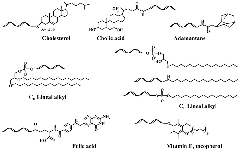 Figure 1. Structure of several lipid-oligonucleotide conjugates.1
Lipid-ASO Conjugation Overview
Direct conjugation creates one molecular construct
In a direct conjugate, cholesterol, a fatty acid, a tocopherol-like group, a steroidal structure, or another hydrophobic moiety is joined to an ASO through a defined bond. The product can often be described by sequence, lipid identity, attachment site, and linker composition. Its amphiphilic character may promote association with albumin, lipoproteins, membranes, or other biological components. It may also drive self-association above a concentration-dependent threshold. Because the lipid remains part of the test article, molecular identity, aggregate state, and the fate of any cleaved species must be considered together.
Lipid conjugation is not LNP encapsulation
A lipid nanoparticle platform contains multiple formulation components assembled around or with a nucleic-acid payload. The ASO may be encapsulated, electrostatically complexed, or partitioned into an internal phase without being covalently attached to a lipid. In contrast, a lipid-ASO conjugate retains its lipid at the molecular level unless a cleavable linker is processed. The distinction changes manufacturing and analytics: conjugates require confirmation of covalent identity and residual free lipid or ASO, whereas LNP-based ASO encapsulation requires loading, leakage, particle-size, and formulation-composition measurements. Hybrid systems can place a lipid-conjugated ASO inside a particle, but then both molecular and particle states require control.
| Format | ASO-Lipid Relationship | Dominant Design Variable | Primary Analytical Question |
|---|---|---|---|
| Direct lipid-ASO conjugate | One lipid-bearing molecular species | Lipid structure, linker, and attachment site | Is the intact conjugate pure and chemically defined? |
| Lipidated ASO self-assembly | Conjugate molecules form concentration-dependent assemblies | Hydrophobicity and critical aggregation behavior | Which species exists in buffer, serum, and dosing formulation? |
| LNP-encapsulated ASO | ASO partitions within a multicomponent nanoparticle | Formulation composition and mixing process | How much ASO is encapsulated, free, or released? |
| Lipid-ASO loaded into an LNP | Covalent conjugate is also formulated in a particle | Compatibility between conjugate and particle phase | Can molecular and particle attributes be resolved independently? |
How Lipid Identity Rewrites Distribution?
Plasma carriers become part of the delivery route
Hydrophobic conjugates can associate with albumin and circulating lipoproteins. That association may reduce immediate renal filtration, extend plasma residence, and enable transport across endothelial barriers. It can also redirect the conjugate toward tissues expressing lipoprotein receptors or toward clearance pathways used by the carrier particle. Binding is not a fixed property of the lipid alone: phosphorothioate content, linker polarity, formulation, species-specific plasma composition, and concentration all contribute. An exposure increase should therefore be interpreted with protein-binding and intact-conjugate data rather than attributed automatically to a particular receptor.
Chain length, saturation, and topology alter the balance
A longer or more hydrophobic lipid may strengthen plasma association and membrane partitioning, but excessive hydrophobicity can reduce solubility, increase aggregation, complicate purification, or trap material in extracellular or interstitial compartments. Saturated and unsaturated chains differ in packing behavior; sterols present a rigid topology; branched or multivalent lipids change the exposed hydrophobic surface. A lipid that increases whole-tissue ASO concentration may produce only a small intracellular gain if the conjugate remains bound to extracellular carriers. The relevant design objective is productive exposure in the target cell, not maximal hydrophobicity or bulk-organ accumulation.
- Albumin association can prolong circulation and influence transendothelial transport without proving receptor-mediated uptake.
- Lipoprotein binding may create receptor-dependent distribution, but the responsible carrier class can differ across lipid structures and species.
- Membrane insertion can increase cell association while still leaving most ASO within nonproductive endosomal or surface-bound pools.
- Self-assembly can change apparent size and exposure; concentration and biological matrix should be specified for every measurement.
- Renal and hepatic handling remain relevant because cleavage can regenerate an ASO with distribution properties distinct from the intact conjugate.
From Plasma Association to Productive RNA Engagement
Lipid conjugation creates several possible transport routes, but each route contains conditional gates. A useful study separates the fate of the intact amphiphile from the fate of released ASO and measures the cell type in which target RNA is actually modulated.
- The administered conjugate disperses or self-associates in the dosing matrix; aggregation, adsorption, or precipitation can change the effective dose before tissue exposure begins.
- In blood or interstitial fluid, the conjugate partitions among free, albumin-bound, lipoprotein-bound, and other protein-associated states.
- Carrier interactions influence endothelial transfer and tissue residence. Increased interstitial exposure does not establish entry into parenchymal cells.
- Cells internalize the conjugate through adsorptive, receptor-associated, or mixed endocytic routes determined by lipid and ASO chemistry.
- Endosomal processing, linker cleavage, and membrane interactions determine whether an active ASO species reaches cytosolic or nuclear compartments.
- The ASO binds its intended RNA and produces RNase H cleavage, splice modulation, or steric blockade, followed by a mechanism-matched protein or phenotypic response.
Molecular Design of Lipid-ASO Conjugates
| Design Variable | Potential Benefit | Common Failure Mode | Matched Comparison |
|---|---|---|---|
| Lipid hydrophobicity | Stronger protein or membrane association | Poor solubility, aggregation, extracellular retention | Hydrophobicity series with constant ASO and linker |
| Lipid topology | Different carrier and membrane interactions | Unpredictable tissue redistribution | Sterol, linear-chain, and branched variants |
| Spacer polarity and length | Improved solubility and lipid presentation | Excess flexibility or loss of binding behavior | Short and extended hydrophilic spacers |
| Linker stability | Control over active-species release | Premature cleavage or intracellular persistence | Stable and cleavable matched pair |
| Attachment position | Preservation of sequence architecture | Reduced hybridization or altered protein recruitment | 5', 3', and parent ASO variants |
A Lipid Selection Map
The most useful lipid is the one that addresses the limiting biological barrier while preserving a manufacturable and interpretable conjugate. The following map can guide a small comparative panel before resources are committed to extensive in vivo screening.
| Research Objective | Lipid Hypothesis | Early Readout | Decision Boundary |
|---|---|---|---|
| Increase plasma residence | Promote controlled albumin or lipoprotein association | Free fraction, intact exposure, urinary recovery | Longer exposure must not be caused by insoluble aggregates |
| Improve muscle interstitial access | Use carrier association to support endothelial transport | Cell-resolved muscle distribution and splice or RNA effect | Interstitial accumulation is not equivalent to myocyte delivery |
| Increase membrane interaction | Tune hydrophobic surface for cell association | Internalized ASO and subcellular localization | Surface binding must be separated from endocytosis |
| Create receptor-linked uptake | Favor a defined lipoprotein carrier pathway | Competition, receptor perturbation, and RNA modulation | Carrier binding and receptor causality must both be shown |
| Enable intracellular release | Combine lipid transport with a cleavable linker | Intact conjugate and released-ASO time courses | Cleavage must occur after delivery rather than in plasma |
Analytical Challenges of an Amphiphilic Conjugate
Chemical identity is only the first layer
Mass spectrometry and chromatographic methods should confirm sequence, lipid mass, attachment, purity, and removal of unconjugated ASO or free lipid. Reverse-phase retention can distinguish a hydrophobic conjugate, but strong adsorption or aggregate formation can distort recovery. Orthogonal ion-exchange or capillary methods can help resolve charge-related impurities. Stability assays should track linker cleavage, ASO shortening, oxidation, lipid degradation, and loss to container surfaces. Concentration measurements need an extraction or disruption procedure validated for the amphiphilic state rather than borrowed directly from an unconjugated ASO assay.
Physical state changes with matrix and concentration
Dynamic light scattering, size-exclusion methods, ultracentrifugation, microscopy, or other approaches can probe self-association, but no single method defines every species. Measurements should be performed in formulation, after dilution, and in serum-containing matrices because a preparation that appears monomeric in buffer can bind proteins or form assemblies after dosing. Critical micelle-like transitions, colloidal size, turbidity, and recovery can be evaluated across concentration. Analytical reports should state temperature, buffer, incubation time, and protein content so physical-state results can be reproduced and related to biological assays.
- Confirm molecular mass, sequence, attachment position, lipid identity, and conjugate purity with orthogonal methods.
- Quantify intact conjugate, released ASO, and relevant metabolites rather than reporting total oligonucleotide alone.
- Test adsorption, dilution stability, freeze-thaw behavior, and aggregate formation in the intended container and dosing matrix.
- Measure plasma-protein and lipoprotein partitioning at pharmacologically relevant concentrations and in each study species.
- Retain an identical parent ASO and a lipid-only or linker control where the experimental question requires attribution.
Building a Biological Evidence Chain
Cell studies must distinguish association from activity
Fluorescent intensity can reflect membrane-bound, endosomal, degraded, or intact conjugate. Cell studies should therefore separate surface binding, internalization, subcellular distribution, intact intracellular ASO, target RNA, and target protein. Serum-free conditions may exaggerate membrane insertion, while transfection reagents bypass the delivery question. A mechanism-matched ASO screen should include parent ASO, sequence control, lipid or linker variants, and relevant receptor or carrier perturbations. Productive uptake is demonstrated when a sequence-dependent RNA effect follows the predicted transport mechanism and is not explained by cellular stress.
In vivo studies require cell-resolved exposure
Tissue homogenates cannot distinguish vascular, interstitial, phagocytic-cell, and target-cell material. Fractionation, cell sorting, imaging, or spatial measurements can test whether increased tissue exposure reaches the intended cell population. Pharmacokinetic assays should resolve intact lipid conjugate and released ASO whenever feasible. Dose and time courses can reveal nonlinear protein binding, self-association, carrier saturation, or delayed intracellular activity. Safety readouts should cover liver and kidney effects, hematology, complement or cytokine signals when relevant, and histopathology. Species differences in albumin, lipoproteins, and endothelial transport should be treated as mechanistic variables.
- Advance a candidate only when improved exposure is linked to an RNA endpoint in the intended cell type.
- Reject a distribution claim based solely on whole-organ fluorescence or total ASO concentration.
- Use recovery time points to determine whether prolonged tissue residence produces durable activity or persistent inactive material.
Advantages, Failure Modes, and Safety Boundaries
- Alters distribution without carrier formulation.
- Maintains defined ligand-to-ASO ratio; avoids nanoparticle manufacturing complexity.
- Lipids are synthetically tunable; small series can probe binding, tissue access, and intracellular behavior.
- Lipid may alter ASO protein-binding and clearance unpredictably.
- Bulk accumulation may not translate to productive cell entry.
- Risks: aggregation, precipitation, complement responses, altered renal/hepatic clearance, linker instability, hydrophobic impurities.
- Lipid can reduce activity by interfering with hybridization or ASO mechanism proteins.
- Balance solubility, intact exposure, cell-type delivery, target modulation, and tolerability.
- Do not optimize any single metric in isolation.
- Candidate selection must account for both efficacy and off-target/toxicological liabilities.
Frequently Asked Questions
Q: Is a lipid-conjugated ASO the same as an LNP-formulated ASO?
A: No. A lipid-conjugated ASO contains a covalently attached lipid as part of one molecular construct. An LNP-formulated ASO is associated with a multicomponent particle and may not be chemically linked to any lipid.
Q: Which lipids are commonly explored for direct ASO conjugation?
A: Cholesterol-like structures, long-chain fatty acids, tocopherol-like groups, and other hydrophobic motifs are common starting points. Their performance depends on linker, ASO chemistry, route, dose, and biological context.
Q: Does greater hydrophobicity always improve ASO delivery?
A: No. Greater hydrophobicity can strengthen protein or membrane association, but it can also reduce solubility, increase aggregation, and promote extracellular retention. Productive target-cell activity is the relevant outcome.
Q: Must the lipid be cleaved from the ASO?
A: Not always. Some conjugates remain active while intact, whereas others benefit from intracellular release. Stable and cleavable matched variants can determine whether release is required for a particular design.
Q: How can plasma-carrier involvement be tested?
A: Measure partitioning among free, albumin-bound, and lipoprotein-associated states, then combine carrier depletion, competition, receptor perturbation, and intact-conjugate pharmacokinetics with RNA activity.
Q: What controls are essential in a lipid-ASO study?
A: Include the identical parent ASO, a sequence-control conjugate, lipid or linker variants, and relevant carrier or receptor controls. Cell studies should distinguish surface binding, internalization, and productive RNA modulation.
Published Data
Case 1: Lipophilic Compound-Conjugated ASOs for Exon Skipping in Duchenne Muscular Dystrophy
This study evaluates antisense oligonucleotides (ASOs) conjugated with lipophilic compounds to enhance exon skipping in Duchenne muscular dystrophy (DMD). Researchers synthesized ASO conjugates using bile acids (UDCA, HDCA, TUDCA) and omega-3 fatty acids (DHA, EPA), assessing their physicochemical properties and in vitro biological activity. Unmodified ASOs primarily exist as 2 nm monomers in aqueous solution; however, 5'-end conjugation with ursodeoxycholic acid (UDCA) drives supramolecular self-assembly into ~150 nm nano-aggregates, while dual-end conjugation yields even larger structures. This lipophilic conjugation strategy leverages neutral lipids—such as fatty acids, cholesterol, and squalene—to modulate ASO supramolecular assembly, overcome cellular membrane barriers, and enhance functional delivery and exon-skipping potency in skeletal and cardiac muscle tissues.
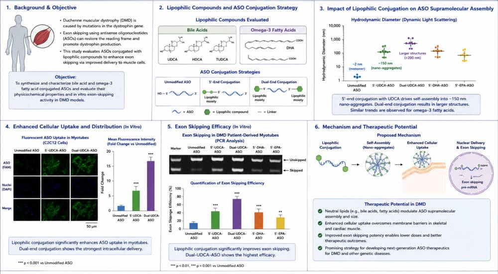 Figure 2. Lipophilic ASO conjugates for DMD exon skipping.
Overview of What Creative Biolabs Can Provide
Creative Biolabs can support a lipid-ASO program from parent-sequence preparation and lipid-linker design through conjugate characterization, formulation comparison, cell-based testing, and in vivo evaluation. The services below are selected from the Gene Therapy Services branch and correspond to distinct questions raised in this resource.
| Research Need | Related Creative Biolabs Support | How It Connects to the Current Resource Topic |
|---|---|---|
| Develop a direct lipid-ASO conjugate | Lipid-Conjugated Antisense Oligonucleotide (ASO) Development Service | Integrates lipid selection, linker and attachment-site design, conjugation, purification, and characterization. |
| Compare lipid with other ligand formats | Antisense Oligonucleotide (ASO) Conjugate Development Services | Places lipid conjugation alongside GalNAc, peptide, antibody, polymer, aptamer, and small-molecule strategies. |
| Define an ASO delivery strategy | Antisense Oligonucleotide (ASO) Delivery Services | Matches the intended tissue, cell type, route, and RNA mechanism with an appropriate delivery approach. |
| Evaluate LNP encapsulation as an alternative | Custom LNP based Antisense Oligonucleotide (ASO) Encapsulation Service | Provides a particle-based comparator when shielding and high cargo loading are more important than direct conjugation. |
| Prepare a conjugate-ready ASO series | Custom Antisense Oligonucleotide Synthesis | Generates matched parent, control, functionalized, and chemistry variants for lipid and linker comparisons. |
| Screen lipid-ASO activity in vitro | Antisense Oligonucleotide (ASO) In Vitro Screening Service | Tests free uptake, concentration response, RNA modulation, cellular stress, and mechanism-specific controls. |
| Assess distribution and pharmacology in vivo | In Vivo Study Service for Antisense Therapeutics | Connects intact exposure, tissue and cell distribution, target modulation, and tolerability. |
To discuss a lipid structure, linker, or comparison strategy tailored to your ASO program, contact us today to connect with our scientific team.
References
- Fàbrega C, Aviñó A, Navarro N, et al. Lipid and peptide-oligonucleotide conjugates for therapeutic purposes: from simple hybrids to complex multifunctional assemblies. Pharmaceutics, 2023, 15(2): 320. https://doi.org/10.3390/pharmaceutics15020320 Distributed under Open Access license CC BY 4.0, with modification.
- Marchesi E, Cortesi R, Preti L, et al. Antisense oligonucleotides conjugated with lipophilic compounds: Synthesis and in vitro evaluation of exon skipping in Duchenne muscular dystrophy. International Journal of Molecular Sciences, 2022, 23(8): 4270. https://doi.org/10.3390/ijms23084270
