Sialyl-Lewis A vs. Sialyl-Lewis X: A Guide to Specificity in Cancer Research
As a team of biological specialists at Creative Biolabs, we have dedicated our careers to the complex and fascinating world of glycobiology. In our work supporting research projects, two molecules repeatedly emerge as critical targets in oncology: sialyl-Lewis A (sLeA) and sialyl-Lewis X (sLeX). To a researcher, these two glycans can be the source of significant frustration. They are structurally similar, often coexist in the same tissues, and are notoriously difficult to distinguish. An antibody that cross-reacts can derail an entire study, leading to false positives, inaccurate quantification, and incorrect conclusions. This challenge is precisely where our expertise comes into play. We specialize in developing highly specific tools to solve this exact problem. Our services, such as the Custom Anti-sLeA (CA19-9) Antibody Development for Pancreatic Cancer Research and the Custom Anti-sLeX Antibody Development for Cancer Metastasis Research, are built on a foundation of rigorous, multi-platform screening to ensure absolute specificity. There is a guide for scientists navigating this challenge. We will directly compare sLeA and sLeX, clarify their distinct roles in cancer, and explain why specificity is non-negotiable for advancing your research.
What Are Sialyl-Lewis A (sLeA) and Sialyl-Lewis X (sLeX)?
At the most basic level, sLeA and sLeX are complex glycans. They are classified as tetrasaccharides, meaning each one is built from a specific sequence of four sugar molecules. Both sLeA and sLeX are built from the same four components:
- N-acetylneuraminic acid (Neu5Ac)
- Galactose (Gal)
- Fucose (Fuc)
- N-acetylglucosamine (GlcNAc)
The Critical Structural Difference
The core of this difference lies in the glycosidic bonds, the chemical links that connect the sugar units.
- Sialyl-Lewis A (sLeA) is built on a Type 1 Chain (Galβ1-3GlcNAc). The fucose is attached in an α1-4 linkage to the GlcNAc.
- Sialyl-Lewis X (sLeX) is built on a Type 2 Chain (Galβ1-4GlcNAc). The fucose is attached in an α1-3 linkage to the GlcNAc.
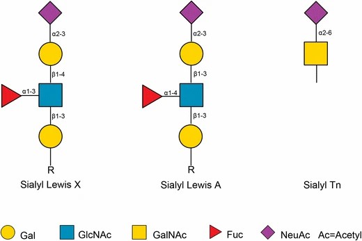 Fig.1 Structural comparison of sLeA, sLeX, and sTn.1
This may seem like a minor chemical detail, but it has massive biological consequences. This shift in linkage changes the three-dimensional shape of the molecule. This new shape, in turn, dictates which proteins each glycan can bind to. Here is a direct comparison to help visualize the difference:
| Feature | Sialyl-Lewis A (sLeA) | Sialyl-Lewis X (sLeX) |
|---|---|---|
| Full Structure | Neu5Acα2-3Galβ1-3[Fucα1-4]GlcNAc | Neu5Acα2-3Galβ1-4[Fucα1-3]GlcNAc |
| Core Chain | Type 1 (Galβ1-3GlcNAc) | Type 2 (Galβ1-4GlcNAc) |
| Fucose Linkage | α1-4 to GlcNAc | α1-3 to GlcNAc |
| Common Name | CA19-9 | CD15s |
| Primary Role | Tumor Marker (Secretion) | Adhesion Ligand (Metastasis) |
Sialyl-Lewis A vs. Sialyl-Lewis X: Roles in Cancer
The Role of Sialyl-Lewis A (CA19-9) in Cancer
When clinicians and researchers talk about Sialyl-Lewis A, they are often using its clinical name: CA19-9 (Carbohydrate Antigen 19-9). In gastrointestinal cancer cells, the enzymes that build sLeA are overexpressed. The glycan is attached to large proteins called mucins (like MUC1 and MUC16), which are then shed from the tumor surface into the bloodstream. This shedding is what allows it to be measured with a simple blood test. While it is the standard for pancreatic cancer, elevated levels of CA19-9 can also be found in other conditions, including other gastrointestinal cancers (like colorectal and gastric cancer) and some benign conditions (like pancreatitis and cirrhosis). CA19-9 is one of the most widely used and FDA-approved serological biomarkers in oncology. Its primary application is in the management of pancreatic cancer. It is used to aid in the diagnosis of pancreatic adenocarcinoma in symptomatic patients and to detect disease recurrence after surgery or treatment.
The Role of Sialyl-Lewis X (sLeX) in Cancer
At Creative Biolabs, we focus on the critical differences between cancer glycans, and sialyl-Lewis X (sLeX) is a major target. Unlike the shed marker sLeA, we understand sLeX as a functional driver of cancer progression. Its role is direct and mechanical: it is the key tool cancer cells use to initiate metastasis, or the spread to distant organs. This process isn't random. It begins when a circulating tumor cell must adhere to a blood vessel wall. Aggressive cancer cells achieve this by overexpressing sLeX, creating a sticky surface. This sLeX then binds specifically to selectin proteins on the vessel walls. This binding is the crucial step that allows the cell to slow down, roll, and stick firmly. Once attached, the cell can exit the bloodstream to form a new tumor. Therefore, high sLeX expression is a direct indicator of metastatic potential, making it a vital target for research.
The Critical Need for sLeA and sLeX Specificity in Research
This specificity challenge between sLeA and sLeX is the most critical research problem. We've seen many researchers struggle with confusing data that traces back to this one issue. Imagine you are in the middle of an ELISA kit development project to quantify CA19-9 for pancreatic cancer monitoring precisely. If your antibody cross-reacts with sLeX, your assay's background will be high and your measurements inaccurate, especially in inflammatory conditions where sLeX is also present. The reverse is even more critical. Consider a therapeutic program designed to block metastasis by targeting sLeX. If your antibody isn't perfectly specific, it will also bind to the large amounts of sLeA shed by gastrointestinal cancer cells. This means your expensive antibody drug will be "used up" by the wrong, soluble target in the bloodstream before it can even reach the tumor's surface, causing your in vivo models to fail. This is why having two distinct, ultra-specific antibodies is the essential foundation for success.
- For sLeA Research: You need a specific antibody (like our anti-sLeA antibodies) to ensure your assay quantifies only CA19-9 without false positives from sLeX.
- For sLeX Research: You need a specific antibody (like our anti-sLeX antibodies) to ensure your functional assays and therapeutic candidates are only targeting the metastatic sLeX determinant.
Our Solutions: Your Partner in Glycan Specificity
At Creative Biolabs, we have built our reputation on tackling these exact specificity challenges. We are a dedicated research partner. We provide end-to-end solutions for scientists working on sLeA, sLeX, and other complex glycan targets. We understand that your research depends on tools you can trust.
Custom Anti-sLeA (CA19-9) Antibody Development for Pancreatic Cancer Research
Target the gold standard biomarker for pancreatic cancer. We assist researchers in developing custom monoclonal or polyclonal antibodies that are highly specific for the sialyl-Lewis A (CA19-9) antigen. We use cutting-edge screening platforms, including comprehensive glycan arrays, to ensure your antibody has minimal to zero cross-reactivity with sLeX, Lewis A, and other related structures. This is ideal for developing next-generation diagnostic assays, research reagents for IHC/IF, and tools for studying gastrointestinal cancer biology.
Custom Anti-sLeX Antibody Development for Cancer Metastasis Research
Target the engine of metastasis. Our scientific team will partner with you to design, produce, and validate high-affinity antibodies specific to the sLeX determinant. We rigorously test all clones against sLeA and other fucosylated glycans to guarantee specificity. This validation is essential for functional studies. These antibodies are critical tools for studying selectin-mediated cell adhesion, tumor cell rolling, metastatic potential, and for use as potential therapeutic blockers.
In cancer research, details matter. sialyl-Lewis A and sialyl-Lewis X are not interchangeable. Their structural similarity is a fundamental challenge that can invalidate research. The future of the field lies in tools that can not only reliably distinguish these two isomers but also identify the specific proteins that carry them. Don't let cross-reactivity compromise your data. Contact our glycan experts today, and let's build the specific, validated tools you need to make the next breakthrough.
FAQs
How does Creative Biolabs validate that your anti-sLeX antibody is truly specific and won't cross-react with sLeA?
This is our top priority. We use comprehensive glycan array screening as our gold standard. We test all antibody clones against a wide panel of related structures, including sLeA, Lewis A, Lewis X, and other fucosylated glycans. We only advance clones that demonstrate exceptionally high affinity for the target and zero detectable cross-reactivity with these critical off-targets.
Do you provide the antibodies, or can you help us develop a complete quantitative ELISA kit?
We do both! We can deliver a highly validated antibody for your own assays, but our expertise shines in end-to-end ELISA kit development. We can build a complete, optimized, and ready-to-use sandwich ELISA kit based on your specific requirements, helping you get reliable quantitative data faster.
What other glycan targets beyond sLeA and sLeX do you have experience with?
Our expertise covers a vast range of cancer-associated carbohydrate antigens. Beyond sLeA and sLeX, we have extensive experience developing high-specificity antibodies against the Tn, sialyl-Tn (sTn), and T antigens. We are fully equipped to tackle virtually any complex glycan target your research requires.
What information do I need to provide to start a custom anti-glycan antibody project with you?
Starting is simple. All we need is your target glycan structure (like sLeA or sLeX) and your intended application (like ELISA, IHC, or FC). Our scientific team will then consult with you to design the best antigen, screening strategy, and validation plan to ensure the final antibody meets your exact research needs.
Reference:
- Zhang, Zejian, Manfred Wuhrer, and Stephanie Holst. "Serum sialylation changes in cancer." Glycoconjugate journal 35.2 (2018): 139-160. Distributed under Open Access license CC BY 4.0, without modification. https://doi.org/10.1007/s10719-018-9820-0
Supports
- Sulfatide and Anti-Sulfatide Antibodies Overview
- TACAs Overview
- Guide to Blood Group Antigens
- Comparing sLeA and sLeX Roles in Cancer
- CA19-9 as a Pancreatic Cancer Biomarker
- Lewis Antigen System Overview
- TACA-Targeted ADCs, CAR-Ts, and RICs