The quest for background-free medical imaging has led to significant breakthroughs in 19F Magnetic Resonance Imaging (MRI), yet the challenge of sensitivity remains a critical hurdle for pharmaceutical developers. By harnessing the unique physicochemical properties of liposomal carriers, researchers can now entrap specific anions to dramatically enhance contrast signals without compromising biocompatibility. Creative Biolabs stands at the forefront of this innovation, leveraging decades of expertise in lipid-based drug delivery to translate these complex physicochemical phenomena into robust, commercially viable imaging and theranostic agents for our global clients.
Developing effective contrast agents for 19F MRI requires overcoming the inherent low sensitivity of the modality while ensuring the carrier system remains stable in biological environments. The entrapment of fluoride (F⁻) and hexafluorophosphate (PF₆⁻) anions within liposome inner cavities offers a promising solution, altering magnetic resonance properties to enable precise monitoring of drug delivery systems. This approach not only addresses the need for improved MRI contrast agents but also opens new avenues for tracking biodistribution in real-time. Creative Biolabs provides the formulation expertise and analytical precision necessary to turn these promising research concepts into optimized, reproducible deliverables for your pipeline.
To understand the potential of this technology, it is essential to grasp the underlying principles governing the interaction between encapsulated ions and the lipid bilayer.
The 19F MRI Advantage
Unlike traditional proton (1H) MRI, which contends with a high background signal from water in the body, 19F MRI offers a "hot spot" imaging capability.
The Compartmentalization Effect
The physical confinement of ions within a nanocarrier changes their behavior.
Recent experimental analyses have shed light on how specific formulation parameters, such as anion type and vesicle size, can be manipulated to maximize imaging sensitivity. The following findings highlight the critical design rules for next-generation contrast agents.
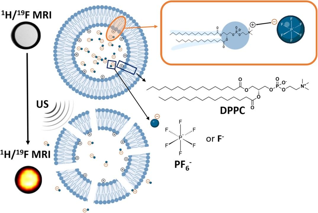 Fig. 1 Schematic representation of the hypothesized interactions between PF6-/F- and the phospholipid bilayer. 1
This research aimed to quantify how the encapsulation of different anions alters magnetic resonance properties compared to their free state. By measuring the Longitudinal (T1) and Transverse (T2) relaxation times of Fluoride (F⁻) and Hexafluorophosphate (PF6−), the study revealed a distinct "Compartmentalization Effect." The results demonstrated that PF6− induces a dramatic two-order magnitude decrease in T1 relaxation times, outperforming F⁻. This enhancement is attributed to the larger, more hydrophobic nature of PF6−, which facilitates stronger dynamic interactions with the lipid bilayer, making it a superior candidate for high-sensitivity "MRI reporters."
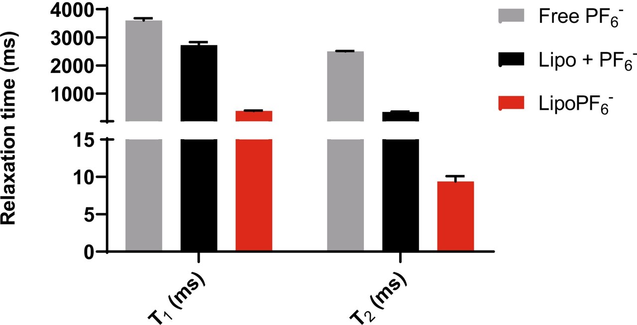 Fig. 2 19F longitudinal (T1) and transverse (T2) relaxation times of liposomes encapsulating 150 mM of PF6−. 1
To understand the physical drivers of contrast sensitivity, researchers analyzed the correlation between liposome diameter and relaxation rates. The study found a strong inverse relationship: relaxation enhancement is substantially more pronounced in smaller vesicles (e.g., <100 nm). This phenomenon occurs because smaller liposomes possess a higher surface-area-to-volume ratio, increasing the probability of entrapped anions interacting with the membrane surface (the site of relaxation enhancement). Consequently, precise control over vesicle size is identified as a critical functional parameter for maximizing MRI signal intensity.
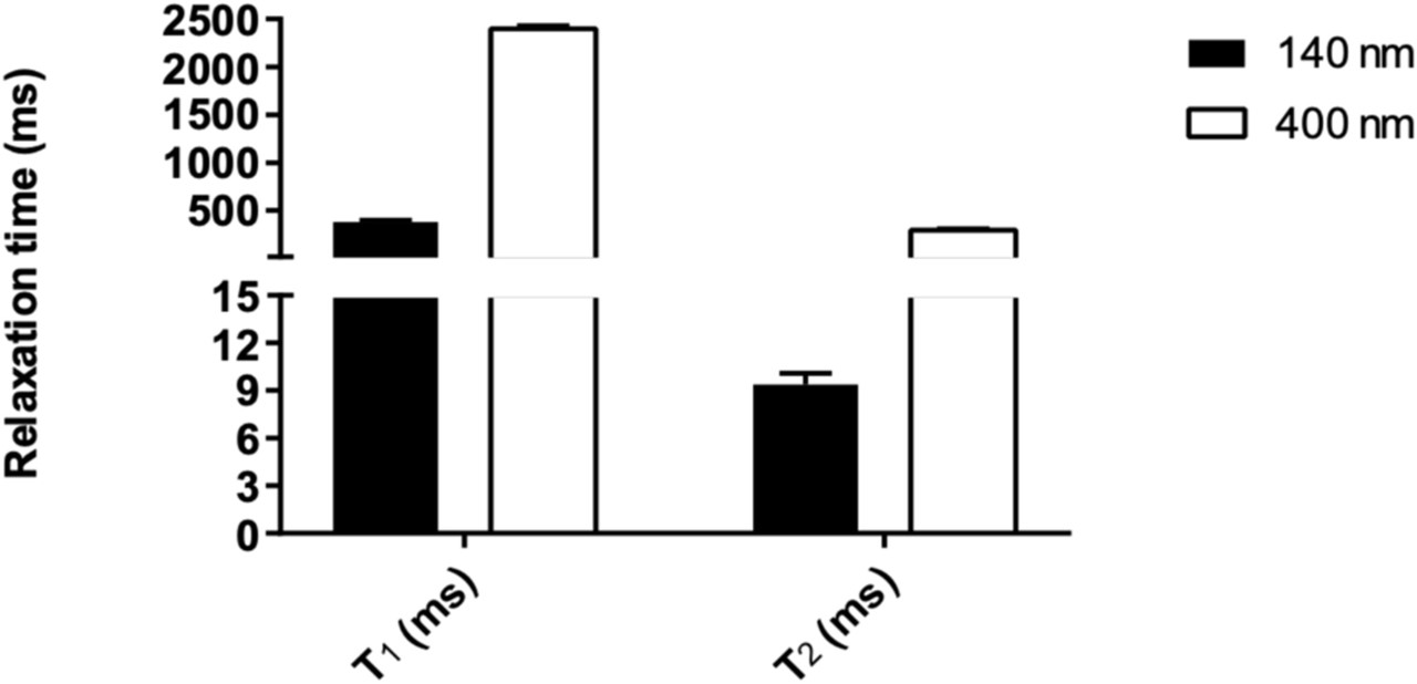 Fig. 3 19F longitudinal (T1) and transverse (T2) relaxation times of PF6−-encapsulating liposomes. 1
To validate the potential for tracking payload delivery, the study investigated the reversibility of the relaxation enhancement upon membrane disruption, a important aspect for designing "smart" theranostic agents. By monitoring T1 relaxation times before and after the addition of a surfactant to lyse the liposomes (simulating drug release), researchers observed a distinct "contrast switch." Upon vesicle rupture and anion release into the bulk medium, the T1 relaxation times reverted to their long, native values similar to free solution. This reversible magnetic signature provides a powerful mechanism to distinguish between the intact drug carrier and the released payload, enabling precise, real-time monitoring of drug bioavailability and biodistribution.
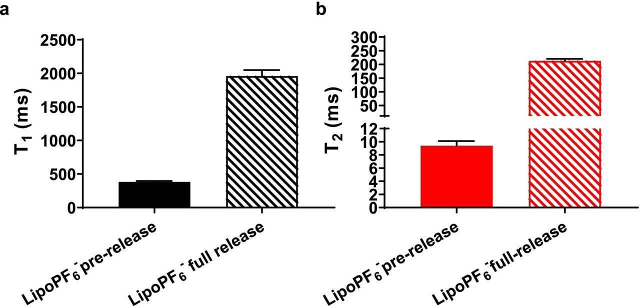 Fig. 4 19F longitudinal (a) and transverse (b) relaxation times of LipoPF6− before and after 100% US-induced content release. 1
The transition from a theoretical concept to a viable diagnostic tool requires more than just raw materials, it demands expertise. At Creative Biolabs, we are ready to partner with you to overcome the formulation challenges inherent in high-sensitivity MRI contrast agents. Reach out to our scientific team to discover how our tailored solutions can accelerate your research in Liposomal F− and PF6− Anion Impact and advanced lipid delivery systems.
Creative Biolabs offers a fully integrated suite of services designed to accelerate your research in anion-based liposomal contrast agents. From initial formulation to rigorous in vivo validation, our team ensures your specific project needs are met with precision.
| Services/Products | Description | Inquiry |
|---|---|---|
| Liposome Development | Custom thin-film hydration and microfluidic encapsulation for hydrophobic compounds. | Inquiry |
| Advanced Characterization | Comprehensive analysis including Size, PDI, Zeta Potential, and Drug Loading efficiency. | Inquiry |
| Process Optimization | Refining formulation parameters to maximize stability and encapsulation rates. | Inquiry |
Reference
 For Research Use Only. Not For Clinical Use
Supports
Online Inquiry
Creative Biolabs is a world-leading liposome provider with extensive experience in liposome preparation, custom formulation development, and characterization.

© 2026 Creative Biolabs. All rights reserved.