Liposome Impact on Membrane Property
Introduction Research Insights Products & Services Resources
The effective delivery of therapeutic agents into the intracellular space is a pivotal challenge in modern biopharmaceuticals. While the potential for gene therapies and intracellular drug targets is vast, the plasma membrane remains a formidable gatekeeper. At Creative Biolabs, we specialize in navigating this complex biological landscape. By leveraging advanced lipid-based drug delivery systems, we empower researchers to bypass traditional barriers. Our expertise in formulation and characterization ensures that your therapeutic cargo reaches its destination, maintaining both high delivery rates and cellular viability.
The Intracellular Access Dilemma
In the pursuit of next-generation therapies, researchers face a critical bottleneck: the plasma membrane prevents most diagnostic and therapeutic agents from reaching their intracellular targets.
Standard delivery methods often rely on endocytosis, a slow process that frequently traps cargo in lysosomes, leading to degradation before therapeutic effect. Lipid-based drug delivery systems (LBDDS) offer a promising solution to this dilemma. By utilizing advanced cationic carriers, we can facilitate direct entry into the cytosol. Creative Biolabs provides the specialized knowledge and technological platform required to translate these complex lipid interactions into robust, scalable delivery solutions for your specific research needs in intracellular delivery.
The Barriers to Entry
The plasma membrane functions as a highly selective barrier, rigorously excluding exogenous entities. Conventional delivery vectors face significant physicochemical obstacles:
-
Steric and Size-Dependent Exclusion: Large macromolecular complexes are typically barred from passive entry due to size constraints.
-
Electrostatic Repulsion: The net negative surface charge of the plasma membrane (glycocalyx) effectively repels neutral or anionic particles, preventing adherence.
-
Endosomal Entrapment: Cargo internalized via standard endocytic pathways is frequently sequestered within endosomes, leading to degradation in the lysosomal compartment before cytosolic release can occur.
The Cationic Solution
Cationic liposomes circumvent these barriers by leveraging electrostatic adsorption to increase cell surface residence time and fusogenic membrane destabilization to facilitate direct cytosolic entry.
|
Mechanism
|
Description
|
Benefit
|
|
Electrostatic Interaction
|
Positive liposomes bind to negative cell membranes.
|
Increases residence time and proximity.
|
|
Membrane Fusion
|
Lipid bilayers merge, creating a continuous channel.
|
Direct cytosolic delivery, bypassing lysosomes.
|
|
Helper Lipids
|
Lipids like DOPE induce curvature (fusogenicity).
|
Facilitates the destabilization required for fusion.
|
Recent Advancements: Transient Membrane Alterations
Recent studies have shifted focus from simply "getting in" to understanding "what happens when we get in." High-charge density cationic liposomes have been shown to fuse efficiently with plasma membranes, delivering cargo in seconds. However, this massive influx of foreign lipids induces significant, though manageable, changes in the cell's physical state.
Kinetics of Cytosolic Delivery vs. Endosomal Uptake
Comparative studies utilizing time-resolved fluorescence microscopy have been instrumental in distinguishing fusion mechanisms from traditional uptake. By tracking the entry of labeled cargo into the cytosol versus endosomal compartments, researchers have demonstrated that cationic liposomes with optimized charge density initiate fusion events within seconds of contact. Unlike endocytosis, which shows a gradual accumulation of signal in punctate vesicular structures over minutes to hours, fusion results in a rapid, diffuse cytosolic spread of the cargo. This dataset is significant as it confirms the ability of fusogenic carriers to bypass the rate-limiting and degradative endosomal pathway, validating their use for cargo sensitive to acidic or enzymatic degradation.
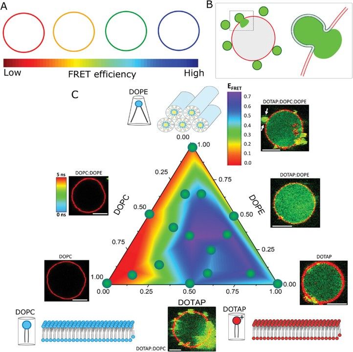 Fig. 1 Fusion efficiency diagram for three-component liposomes. 1
Modulation of Membrane Order and Fluidity
Biophysical assessment of membrane material properties post-fusion, often measured via changes in lipid diffusion coefficients or order parameters, reveals the immediate impact of lipid mixing. Upon fusion, the sudden incorporation of exogenous synthetic lipids results in a measurable perturbation of the native membrane packing. Experimental graphs typically display a shift in lipid mobility, indicating altered fluidity and the creation of local packing defects due to lipid mismatch. Understanding these fluidity shifts is important for formulation, as excessive disordering can compromise cell viability, whereas insufficient modulation may result in failed fusion.
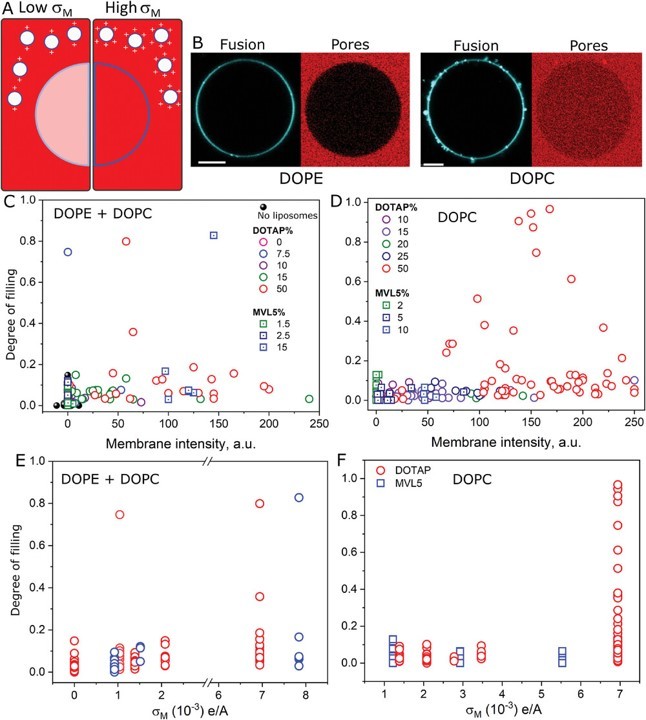 Fig. 2 Charge density determines fusion efficiency and membrane disruption. 1
Induction of Membrane Curvature and Tension Relaxation
Studies focusing on the mechanostructural impact of fusion analyze surface tension changes and curvature induction mediated by helper lipids. Data demonstrates that the insertion of liposomal lipids leads to a quantifiable decrease in plasma membrane surface tension, primarily due to the increased surface area provided by the liposome. Concurrently, the presence of cone-shaped helper lipids induces high local curvature. These mechanical alterations are identified as necessary intermediates for fusion pore formation; however, they highlight the need for precise formulation control to prevent irreversible mechanical damage to the cell.
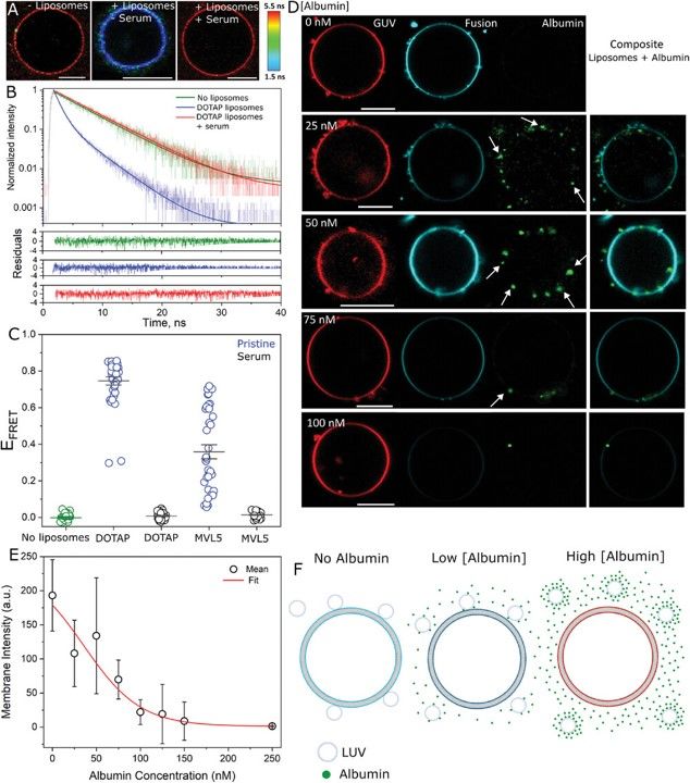 Fig. 3 Protein-coated cationic liposomes are unable to fuse with GUVs. 1
Transient Pore Formation and Cellular Recovery
Electrophysiological or dye-leakage experiments designed to detect non-selective permeability during the fusion window provide critical safety data. Experimental traces often show a spike in non-specific membrane permeability (pore formation) coincident with the fusion event. Crucially, longitudinal data shows a return to baseline impermeability, indicating that these pores are transient and the cell successfully repairs the barrier function. This dataset defines the "therapeutic window," proving that while the delivery mechanism is invasive, the cellular recovery mechanisms are sufficient to restore homeostasis, provided the carrier concentration is within validated limits.
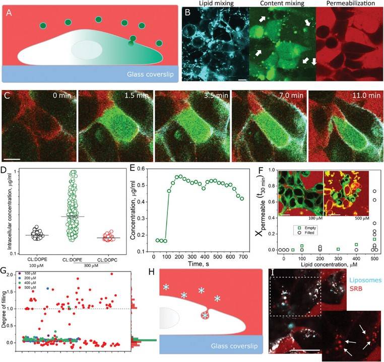 Fig. 4 Intracellular delivery of cargos is associated with plasma membrane permeabilization only at high liposome concentration. 1
Ready to elevate your drug delivery research? At Creative Biolabs, we combine deep scientific expertise with industrial rigor to solve your toughest formulation challenges. Whether you need custom liposome design or advanced characterization of membrane interactions, our team is ready to assist.
Related Services & Products
Creative Biolabs offers a suite of services tailored to the complexities of lipid-based drug delivery. We bridge the gap between biophysical theory and practical application, ensuring your formulations are optimized for both fusion efficiency and cellular safety.
Resources
Reference
-
Hammond, Jayna, et al. "Membrane Fusion‐Based Drug Delivery Liposomes Transiently Modify the Material Properties of Synthetic and Biological Membranes." Small 21.12 (2025): 2408039. https://doi.org/10.1002/smll.202408039. Distributed under Open Access license CC BY 4.0, without modification.

For Research Use Only. Not For Clinical Use