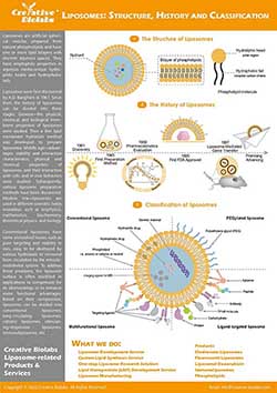
RA & Vitamin D3 Liposome for Skin DC
Introduction Research Insights Products & Services Resources
The shift from broad immunosuppression to antigen-specific tolerance represents the future of autoimmune therapy. By harnessing the body's natural regulatory mechanisms, researchers are developing sophisticated nanocarriers capable of reprogramming the immune system without compromising overall immunity. Creative Biolabs provides the specialized formulation technologies required to bridge these gaps, turning promising research ideas into viable, targeted therapies.
The Challenge & Solution
Developing effective immunotherapies for autoimmune diseases and allergies faces a significant hurdle: delivering therapeutic agents specifically to immune regulators without triggering systemic inflammation. In vivo targeting of dendritic cells (DCs) is particularly complex due to the need for precise uptake and phenotypic control. Lipid-based drug delivery systems offer a promising solution by protecting labile payloads like retinoic acid (RA) and vitamin D3 (VD3) while exploiting natural biological transport pathways.
The Pivotal Role of Dendritic Cells in Tolerance
DCs are the master regulators of the immune system. While they are traditionally known for initiating immune responses against pathogens, they play an equally critical role in maintaining peripheral tolerance. In autoimmune conditions, the goal is to modulate DCs to present antigens in a "tolerogenic" context, thereby instructing T cells to ignore self-tissues rather than attack them.
Why Target the Skin?
The skin is not merely a barrier but an immunologically active organ rich in distinct DC subsets, including Langerhans cells, CD14+ dermal DCs, and CD1a dim dermal DCs. Intradermal administration offers a direct route to these cells, making it an attractive site for vaccination and immune modulation strategies compared to systemic administration.
Lipid Nanocarriers as Tolerogenic Vehicles
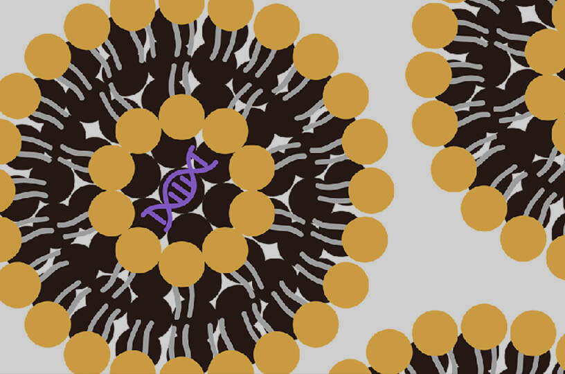
Liposomes, particularly those with specific surface charges (such as anionic phospholipids like DSPG), are ideal candidates for this application.
-
Encapsulation: They can solubilize hydrophobic immunomodulators (RA, VD3).
-
Stability: They protect sensitive cargo from degradation.
-
Targeting: Their physicochemical properties can be tuned to favor uptake by specific DC subsets in the dermis.
Reprogramming Skin Dendritic Cells for Immune Tolerance
Recent investigations have elucidated how specific lipid formulations can differentially affect DC behavior. By utilizing anionic liposomes loaded with tolerogenic adjuvants, researchers have achieved precise modulation of immune pathways.
Selective Migration and Phenotypic Control
Intradermal injection of RA or VD3-loaded DSPG liposomes was found to function as a selective immune modulator, specifically inducing the migration of CD14+ dermal DCs while inhibiting CD1a dim DCs. Importantly, this mobilization does not trigger an inflammatory storm; rather, the migrated CD14+ cells maintain a "partially immature" phenotype. This finding is significant because it demonstrates the ability to engage specific immune subsets without provoking the strong activation typically associated with vaccines, a prerequisite for tolerance.
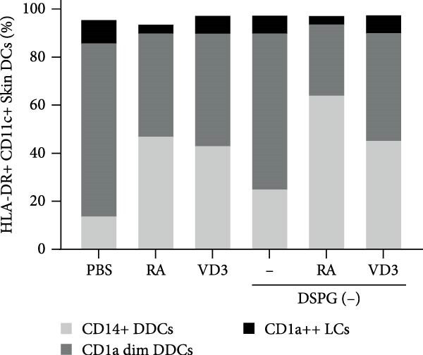 Fig. 1 RA- and VD3-loaded DSPG liposomes Induce migration of CD14+ DDCs from human skin. 1
Induction of Coinhibitory "Brake" Molecules
A key mechanism identified for inducing tolerance is the upregulation of coinhibitory molecules. The study showed that treatment with these liposomes induced the expression of ILT3 in CD1a dim DCs and PD-L1 (specifically by VD3 liposomes) across DC subsets. These molecules act as essential "checkpoints," actively suppressing T cell activation. By equipping DCs with these molecular brakes, the therapy effectively halts the progression of autoimmune responses at the cellular level.
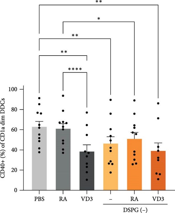 Fig. 2 RA and VD3 liposome injection reduce expression of activation markers and induce expression of tolerogenic markers in mature CD1a dim DDCs. 1
Generation of Functional Regulatory T Cells
The ultimate validation of this approach lies in its ability to influence T cell behavior. The research confirmed that DCs modified by RA/VD3 liposomes successfully differentiated naïve CD4+ T cells into FoxP3+ Tregs with a CD127 low, ICOS+ phenotype. This conversion is critical, as it confirms that the liposomal treatment establishes a complete tolerogenic loop—from targeted delivery in the skin to the generation of functional suppressive T cells—offering a viable pathway for treating allergic and autoimmune disorders.
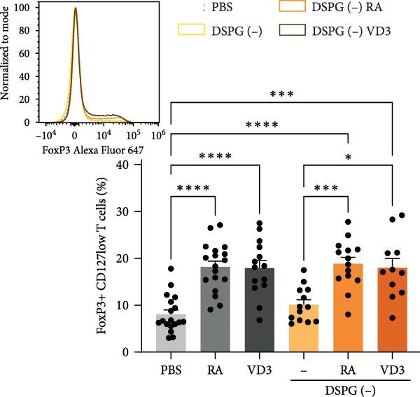 Fig. 3 RA- or VD3-liposome-treated skin DCs induce FoxP3+ CD127low and ICOS+ Tregs expressing functional Treg markers. 1
Are you developing novel immunotherapies for autoimmune diseases or allergies? Do not let formulation challenges hinder your progress. Contact our expert team today to discuss how we can support your specific project needs in skin DC targeting and lipid-based drug delivery. From custom liposome synthesis to validation assays, we are your partners in innovation.
Related Services & Products
Creative Biolabs offers a comprehensive suite of services designed to support the development of tolerogenic lipid nanoparticles. From the initial encapsulation of hydrophobic agents like RA and Vitamin D3 to the complex characterization of anionic liposomes, our team ensures your vectors are optimized for skin DC targeting.
Resources
Reference
-
Nagy, Noémi Anna, et al. "Intradermally Administered Retinoic Acid or Vitamin D3‐Loaded Liposomes Induce Tolerogenic Skin Dendritic Cells." Journal of Immunology Research 2025.1 (2025): 2208155. https://doi.org/10.1155/jimr/2208155. Distributed under Open Access license CC BY 4.0, without modification.

For Research Use Only. Not For Clinical Use