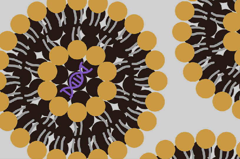
Niosome loaded Zinc Oxide Nanoparticle
Zinc Oxide Nanoparticles Niosomes Research Insights Products & Services Resources
In the development of modern targeted therapies, effectively protecting active pharmaceutical ingredients, overcoming physiological barriers, and precisely targeting diseased tissues remain core challenges for both academia and industry. Recently, the innovative architecture combining inorganic nanoparticles with non-ionic surfactant vesicles (niosomes) has unlocked unprecedented potential for targeted therapies. This page explores the dynamic synergy between green-synthesized zinc oxide (ZnO) nanoparticles and advanced niosomal delivery systems. Creative Biolabs, leveraging deep expertise in lipid-based and nanoparticle delivery technologies, is dedicated to providing top-tier R&D support for global clients, helping you translate these scientific breakthroughs into clinical realities with high commercial value.
The Rise of Green-Synthesized Zinc Oxide Nanoparticles
Traditional chemical synthesis of inorganic nanoparticles often relies on toxic precursors, posing a significant risk for biomedical applications. In recent years, the "green synthesis" strategy, utilizing plant extracts or biological agents, has become a mainstream approach.
-
Core Advantage: Drastically reduces environmental impact while conferring extremely low inherent toxicity to the nanoparticles.
-
Scientific Significance: Provides a highly biocompatible nanomaterial foundation for sensitive cellular interactions and subsequent clinical translation.
Niosomes: The Next-Generation Drug Carrier
Niosomes are vesicular carriers formed by the self-assembly of non-ionic surfactants and cholesterol. While structurally similar to traditional liposomes, they exhibit superior characteristics for industrial applications.
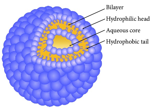 Fig. 1 Structure of niosomes. 1,3
-
Structural Advantage: Possess a unique amphiphilic architecture capable of efficiently encapsulating both hydrophilic and lipophilic drugs simultaneously.
-
Application Focus: Compared to conventional liposomes, niosomes offer higher chemical stability and lower production costs, making them an ideal choice for large-scale industrial manufacturing.
Synergistic Therapeutic Potential of Nanoparticles & Vesicles
When bioactive metal nanoparticles are encapsulated within a niosomal matrix, their therapeutic efficacy is exponentially amplified. The vesicle acts as a biological "Trojan horse," ensuring that the active payload is not prematurely degraded before reaching target sites, such as the tumor microenvironment or stubborn biofilms. Establishes a solid technological pathway for developing next-generation antimicrobials and targeted oncology therapies with broadened therapeutic windows.
Advanced Morphological & Structural Characterization
Confirming the successful synthesis of nanoparticles and their precise encapsulation within vesicles is the primary step in formulation development. Through analytical techniques such as Scanning Electron Microscopy (SEM), Dynamic Light Scattering (DLS), Fourier Transform Infrared Spectroscopy (FTIR), and X-Ray Diffraction (XRD), the morphology and crystalline structure can be accurately mapped. In an ideal optimized model, circular green-synthesized nanoparticles (averaging ~27.6 nm) are stably nested within niosomal vesicles (averaging ~256 nm).
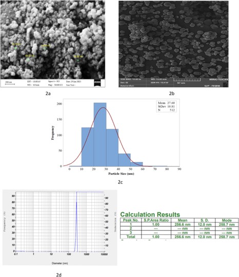 Fig. 2 Characterization of zinc oxide nanoparticles (Zn-NPs). 2,3
Encapsulation Efficiency & Precision Release Kinetics
The core value of a vesicular carrier lies in its ability to securely lock the active payload and release it on demand. Evaluating the encapsulation efficiency (EE%) and in vitro dissolution profile is crucial for determining the success of the delivery system. High-performance niosomal formulations achieve excellent drug loading (e.g., >31% EE) and demonstrate a highly controlled, sustained-release curve—releasing only about 26% of the nanoparticles within 24 hours, compared to a near 99% release rate for free nanoparticles over the same period. Such analyses correspond to the cumulative drug release profiles crucial in pharmacokinetics.
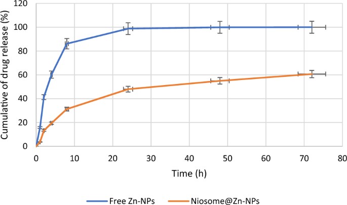 Fig. 3 Release of Zn-NPs from niosomes compared with free form. 2,3
Amplified Antimicrobial & Anti-Biofilm Activity
Bacterial resistance and biofilm formation represent major challenges in modern medicine. Through rigorous microbial inhibition zone assays and biofilm gene expression evaluations, niosome-loaded nanoparticles demonstrate overwhelming superiority. Thanks to the controlled release mechanism, the drug penetrates deeper into biofilms, boosting antimicrobial and anti-biofilm effects by 2 to 4 times compared to free nanoparticles. This evaluation is typically presented via microbial growth inhibition curves and biofilm mass reduction charts.
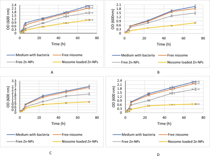 Fig. 4 The antimicrobial effects of niosomes containing Zn-NPs and free Zn-NPs and free niosomes. 2,3
Superior Cytocompatibility & Targeted Anticancer Mechanisms
A successful drug delivery system must strike a perfect balance between "high toxicity to target cells" and "extreme safety for healthy tissues." Testing against normal cell lines (e.g., HEK-293) establishes excellent cytocompatibility. Conversely, when targeting cancer cell lines (such as breast cancer), the niosomal carrier significantly upregulates the expression of apoptosis-related genes (Casp3, Casp9, Bax) while effectively downregulating anti-apoptotic genes (Bcl2). Validation at this biological level is generally reflected in in vitro cell viability (MTT) assay charts and RT-qPCR gene expression heatmaps.
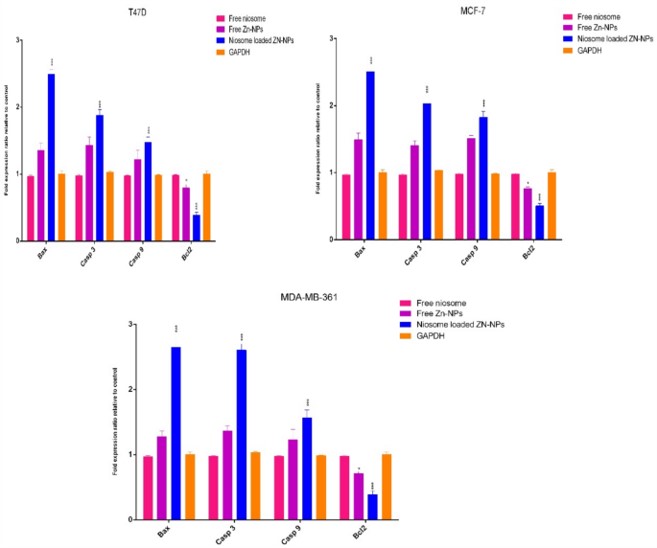 Fig. 5 gene expression in cells exposure to niosome-loaded Zn-NPs, free Zn-NPs, and free niosomes. 2,3
To accelerate your R&D pipeline, Creative Biolabs provides comprehensive, end-to-end solutions for lipid and nanoparticle delivery systems, from conceptual design to industrial scale-up. We offer premium technical services and ready-to-use research products to ensure your experimental data is robust and reproducible.
Related Services & Products
Creative Biolabs boasts industry-leading technology platforms and extensive experience in liposomal and vesicular development. From innovative formulation design and rigorous comprehensive characterization to in-depth biological functional validation, we possess the core competencies required to translate cutting-edge scientific concepts into practical, market-ready deliverables. Choosing Creative Biolabs means partnering with a team of experts equipped with profound scientific heritage and exceptional delivery capabilities. We look forward to collaborating with you to accelerate the clinical transition of your novel drug delivery systems, jointly pioneering the future of nanomedicine.
|
Services/Products
|
Description
|
Inquiry
|
|
Liposome & Niosome Development
|
Tailored design and synthesis of highly optimized non-ionic vesicular carriers specific to your Active Pharmaceutical Ingredient (API).
|
Inquiry
|
|
Process Optimization & Scale-Up
|
Engineering services for the optimization of green synthesis pathways and the industrial scale-up of liposomal encapsulation processes.
|
Inquiry
|
|
High-Purity Lipid Components
|
Pharmaceutical-grade cholesterol and non-ionic surfactants for the in-house formulation of highly stable vesicular carriers.
|
Inquiry
|
Resources
References
-
Ag Seleci, Didem, et al. "Niosomes as nanoparticular drug carriers: fundamentals and recent applications." Journal of nanomaterials 2016.1 (2016): 7372306. https://doi.org/10.1155/2016/7372306.
-
Rezaei, Hossein, et al. "Formulation, preparation of niosome loaded zinc oxide nanoparticles and biological activities." Scientific Reports 14.1 (2024): 16692. https://doi.org/10.1038/s41598-024-67509-5.
-
Distributed under Open Access license CC BY 4.0, without modification.

For Research Use Only. Not For Clinical Use