Anti-Glycan Epitope Mapping Service
Custom Epitope Mapping for Glycan, Glycopeptide, and Site-Dependent Recognition
Creative Biolabs' Anti-Glycan Antibody Research Services integrate target design, glycoform-focused assay selection, and analytical interpretation to support precise anti-glycan epitope mapping. This service is built for researchers who need to identify antibody binding epitopes beyond ordinary peptide mapping, especially when antibody specificity depends on a glycan-only epitope, a glycopeptide neoepitope, or a position-sensitive epitope shaped by where glycosylation is installed.
What This Mapping Service Clarifies
- Whether the binding epitope is driven by carbohydrate alone, by a combined glycan-peptide surface, or by site-specific glycosylation context.
- How far antibody specificity extends across related glycoforms, adjacent glycosylation sites, and truncation series.
- Which assay formats are most suitable for confirming the antibody epitope in a defined research setting.
Background for Anti-Glycan Epitope Mapping
Antibody epitope mapping becomes substantially more complex when the target is not a linear peptide but a carbohydrate-containing determinant. Many anti-glycan antibodies appear selective in one assay yet lose discrimination when tested against related glycans, alternative glycoforms, or the same glycan displayed at a different position on a peptide or protein backbone. This is why a true anti-glycan epitope mapping service must distinguish carbohydrate recognition from scaffold effects rather than treating every project as standard peptide epitope mapping.
In practice, researchers often need to know whether an antibody binds a terminal glycan motif regardless of carrier, whether it recognizes a glycopeptide neoepitope formed jointly by glycan and neighboring residues, or whether the binding event depends on a precise glycosylation site and local sequence geometry. Those three outcomes lead to very different decisions in antibody screening, clone triage, assay development, and downstream reagent positioning.

Fig.1 Overview of distinct approaches for anti-glycan epitope mapping and precise antibody analysis.
- Glycan-only epitopes are defined by carbohydrate features that remain recognizable across multiple carriers.
- Glycopeptide neoepitopes require simultaneous evaluation of glycan identity and the adjacent peptide environment.
- Position-sensitive epitopes demand site-resolved comparison because glycosylation at nearby residues can abolish or reshape antibody binding.
- Reliable mapping improves antibody specificity assessment, counter-screen design, and binder prioritization.
Key Pain Points in Epitope Mapping Service Design
Our anti-glycan epitope mapping service is structured around the practical reasons why antibody mapping projects fail or remain ambiguous.
False Peptide Bias
Some antibodies seem glycan-reactive, yet binding is primarily driven by the backbone or linker environment.
Glycoform Overlap
Closely related structures such as Tn, T, STn, and extended cores can create misleading selectivity claims when comparison panels are narrow.
Site Ambiguity
The same monosaccharide can be recognized at one residue but not at a neighboring residue, especially in mucin-like regions.
Assay Mismatch
A binding epitope identified in one assay format may not reflect the presentation state used in later research workflows.
Orthogonal Panels
We compare glycan series, glycopeptide series, and non-glycosylated controls to identify antibody binding epitopes with better confidence.
Glycan-Only Resolution
Carrier-diverse and structure-related comparisons help determine whether the antibody specificity is carbohydrate-dominant.
Site-Resolved Mapping
Position-swapped and truncation-oriented designs support evaluation of position-sensitive epitopes rather than generic glycan presence.
Interpretive Reporting
Results are summarized as a binding model that clearly separates glycan-driven, glycopeptide-driven, and context-dependent recognition.
Scope of Our Anti-Glycan Epitope Mapping Service
This service is intended for research-stage antibodies, antibody fragments, and related binders when the key question is whether binding is primarily driven by the glycan itself, by a combined glycan-peptide surface, or by local glycosylation context. Project design is customized to the target system, available reagents, and the level of resolution that can be supported experimentally.
Structured Workflow for Custom Epitope Mapping
Each project follows a stepwise design-review-analysis sequence. The exact comparison panel and assay combination are defined case by case, because not every antibody-target pair supports the same level of glycoform, sequence, or site resolution.
Project Definition
Review the binder, target hypothesis, available controls, and the research question that the mapping study must answer.
Panel Design
Select the most informative glycan, glycopeptide, nonglycosylated, or site-variant comparisons that are feasible for the project.
Binding Analysis
Generate comparative binding data across the defined panel and evaluate relative reactivity, selectivity, and context dependence.
Interpretation
Build a data-supported binding model that distinguishes glycan-dominant recognition from glycopeptide- or site-context-dependent recognition.
Reporting
Deliver the study summary, comparative data, observed boundaries of recognition, and recommended next research steps.
Sample Requirements for Antibody Mapping Service
Clear starting information helps determine whether a project is best approached as glycan-focused comparison, glycopeptide neoepitope analysis, or site-context evaluation.

Recommended Submission Information
- Binder identity and format, such as purified antibody, fragment, clone information, or other research-stage reagent details.
- Target background, including the relevant glycan, glycopeptide, glycosylation site hypothesis, or related structures that should be compared.
- Available positive and negative controls, prior screening observations, or internal benchmark data if they exist.
- The primary question to be resolved, for example glycoform selectivity, peptide contribution, site dependence, or cross-reactivity boundaries.
- Any practical constraints that may affect study design, such as sample amount, preferred readout, timeline, or downstream research use.
Project Output and Deliverables
Deliverables focus on what the submitted binder recognized under the tested conditions and where the current data place the boundaries of specificity.
Typical Deliverables
- Project summary describing the study question, panel logic, controls, and any important design constraints.
- Raw and processed comparative binding data generated from the selected test panel.
- A research-use interpretation indicating whether the observed binding pattern is more consistent with glycan-focused, glycopeptide-context-dependent, or site-context-dependent recognition.
- Cross-reactivity observations, study limitations, and practical suggestions for follow-up validation or expanded comparison in later studies.

Need to Identify Antibody Binding Epitopes with Higher Confidence?
If your antibody mapping project involves glycan-dependent selectivity, ambiguous glycopeptide reactivity, or unresolved site effects, our team can design a custom epitope mapping workflow around your exact target question.
Published Data Supporting Glycopeptide Neoepitope Mapping
This open access study is a useful literature example for one important subtype of anti-glycan mapping: a mucin-type O-glycopeptide neoepitope. Wakui et al. showed that the anti-MUC1 antibody SN-101 bound a Tn-modified MUC1 glycopeptide through concurrent contacts with the GalNAc residue and the proximal peptide region, and that SN-101 discriminated Tn-glycosylated from T-glycosylated MUC1 fragments. The same report also showed that additional glycosylation at neighboring residues in the N-terminal Val-Thr-Ser-Ala region could impair recognition. Together, these findings support the value of glycoform-focused and site-context-aware comparison when the target is a glycopeptide rather than a peptide alone.
Why This Literature Example Matters
- It provides a clear example of a glycopeptidic neoepitope in which glycan contact and peptide contact are both required for recognition.
- It demonstrates glycoform discrimination, because SN-101 bound Tn-glycosylated MUC1 but not the corresponding T-glycosylated variant.
- It shows that neighboring glycosylation can alter recognition, supporting the need for site-context comparison in some mucin-like targets.
- It supports the scientific rationale for anti-glycan epitope mapping workflows that do not rely on peptide-only comparison.
- This publication is an illustrative literature case, not a direct validation of every anti-glycan target system.
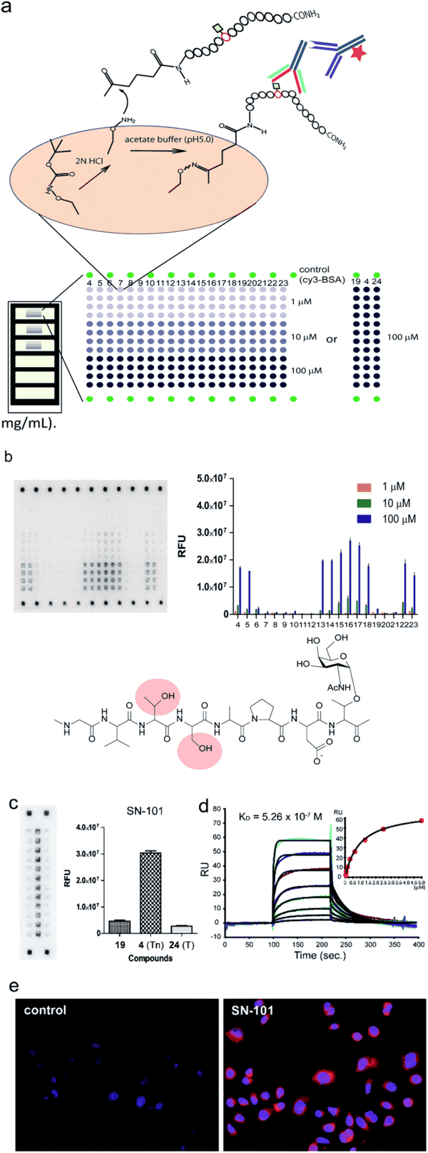
Fig.2 Microarray-based epitope mapping of SN-101 identifies the minimal glycopeptide epitope, shows the impact of neighboring glycosylation, and distinguishes Tn-glycosylated from T-glycosylated MUC1.1
Customer Review
Recommended Products
These product categories can support glycan-focused immunogen preparation, antibody comparison, and research-stage specificity studies.
Carbohydrate Antigen Products
Useful for building defined glycan comparison panels and supporting custom anti-glycan epitope mapping studies with structurally relevant materials.
Learn MoreMonoclonal Antibody Products
Suitable for benchmark comparison, specificity confirmation, and internal reference testing during antibody mapping service projects.
Learn MorePolyclonal Antibody Products
Helpful for exploratory screening and broader reactivity assessment in glycan recognition studies intended for research use.
Learn More