Glycan Microarray-Based Antibody Screening Service
Glycan Microarray-Based Antibody Screening Service
Creative Biolabs profiles antibody glycan specificity and cross-reactivity with
customizable microarrays, matched controls, candidate ranking, and research-focused interpretation for
confident research decisions.
Creative Biolabs works with research teams that need to determine what an antibody
recognizes across structurally related glycans, not simply whether it binds one target preparation. Our glycan
microarray-based antibody screening service is available within Anti-Glycan
Antibody Research Services and can be scoped for broad specificity profiling, target-class panels, clone
comparison, or focused cross-reactivity questions.
The starting point is the research decision that follows the screen. Early discovery may require broad
coverage and conservative hit calling, whereas epitope refinement may need a compact panel that varies one
linkage, branch, terminal group, or presentation feature at a time. Array content, controls, detection format,
and interpretation are therefore aligned with the submitted antibody type and the level of specificity the
project needs to resolve.
What Glycan Microarray Screening Can Resolve
A glycan array places many defined carbohydrate probes in a shared experimental format, allowing relative
binding patterns to be compared under consistent incubation and detection conditions. This is particularly
useful for antibodies because small changes in linkage, branching, sulfation, acetylation, or terminal
substitution may alter recognition even when the nominal glycan family is unchanged.
Motif preference: Identify recurring structural features among positive spots rather than
treating each signal as an isolated hit.
Cross-reactivity boundaries: Compare the intended target with close analogues, related
microbial or mammalian glycans, and negative controls.
Candidate differentiation: Rank clones by signal pattern, reproducibility, breadth, and
selectivity under the same panel conditions.
Presentation sensitivity: Evaluate whether density, linker, carrier, or multivalent display
may be influencing apparent binding.
Service design may begin with a predefined array or a project-defined panel. Creative Biolabs can align array
selection, antibody handling, replicate logic, controls, signal processing, and candidate comparison with the
stated discovery question. Purified antibodies provide the clearest starting material, while selected hybridoma
supernatants, culture supernatants, serum, or plasma may be considered when background and matrix effects can be
controlled.
Specificity and Cross-Reactivity Screening
Specificity screening evaluates the target together with structures that challenge the intended claim.
Depending on the epitope, this may include linkage variants, truncated motifs, stereoisomers, neighboring
substitutions, related glycan classes, carrier-only controls, or biologically relevant off-targets. The
panel is most informative when each comparison has a clear reason for inclusion.
Candidate Comparison and Ranking
Candidate ranking combines signal magnitude with consistency and pattern quality. A high target signal is
less useful when the same clone reacts broadly with related structures. Ranking can therefore incorporate
replicate variation, target-to-near-neighbor separation, off-target burden, concentration response, and the
intended downstream assay or application.
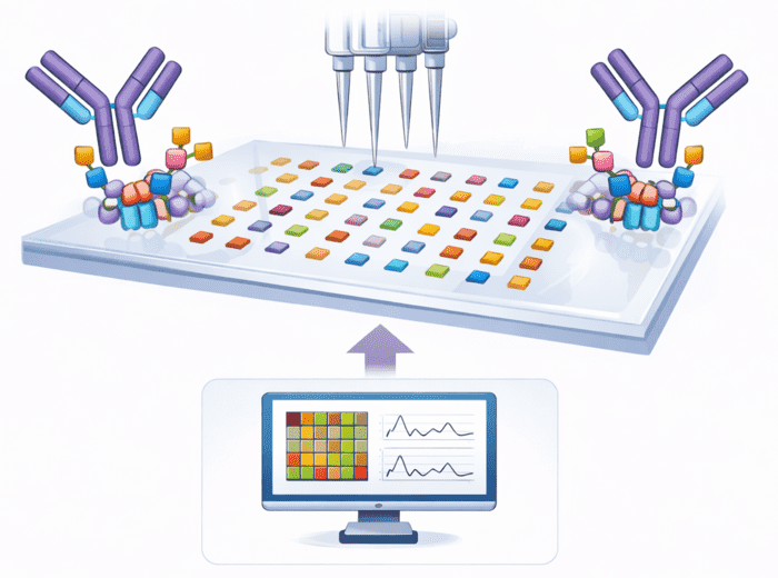
Fig.1 glycan microarray-based screening.
Selecting the Right Glycan Array and Panel
Panel Design Question
What Changes in the Array
Interpretation Benefit
Broad discovery or unknown motif
Diverse glycan classes, broad negative space, multiple presentation contexts
Reveals unexpected binders and broad reactivity, but may require a focused second screen.
Defined target class
Dense structural neighborhood around the target, including linkage and substitution variants
Improves motif-level comparison and supports more defensible selectivity statements.
Clone down-selection
Common panel and matched concentrations for every candidate
Separates clone behavior without confounding from different assay conditions.
Cross-reactivity risk
Project-specific off-targets from host, matrix, pathogen, or process context
Tests the liabilities that matter to the planned research use.
Panel size is not the only determinant of value. Structural coverage, printing chemistry, glycan density, probe
purity, and the relationship between positive and negative controls can matter more than the total spot count. A
targeted panel may be more informative than a broad panel when the decision concerns one narrow epitope
boundary.
Our Screening Workflow
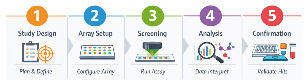
Fig.2 Research workflow for high-throughput glycan array antibody screening.
Define the antibody format, target glycan, relevant near-neighbors, cross-reactivity concerns, and the
decision the screen is intended to support.
Select predefined or custom array content, replicate layout, concentration plan, positive and negative
controls, and appropriate detection reagents.
Prepare compatible samples, incubate the array, apply detection reagents, perform controlled washing,
and acquire spot-level signals.
Perform background correction, assess replicate consistency, normalize when appropriate, identify
binding patterns, and apply conservative hit-calling criteria.
Rank candidates, examine unresolved presentation or cross-reactivity effects, and confirm prioritized
hits using focused arrays, concentration series, or orthogonal assays.
The workflow is adaptable to project findings. A broad initial screen may be followed by a focused array,
concentration series, or independent assay when the data reveal a narrower glycan motif, potential
cross-reactivity, or presentation-dependent binding.
Sample Requirements and Deliverables
Project requirements depend on antibody format, sample purity,
matrix complexity, panel size, and whether the study is exploratory or comparative. Exact amount and
concentration are confirmed after the array and detection route are selected.
Project Input
Sample Requirements
Guides detection chemistry and identifies possible secondary-reagent or Fc-related background.
Supports compatibility review and helps separate sample artifacts from binding behavior.
Defines panel content and the most important specificity comparisons.
Supports matched concentrations, plate layout, replication, and ranking rules.
Clarifies whether the next step is epitope mapping, affinity analysis, assay development, or
native-context testing.
Project Output
Deliverables and Data Interpretation
Array layout and glycan annotation table for the tested panel.
Raw or processed spot images and signal tables, as scoped for the project.
Replicate summaries, background review, and normalized values when normalization is scientifically
appropriate.
Heatmaps, ranked hit lists, clone-comparison plots, or motif-oriented summaries suited to the study
design.
A technical interpretation that separates observed array binding from conclusions that require
orthogonal confirmation.
From Array Hits to Orthogonal Validation
Follow-up testing is selected according to the unresolved question. ELISA-like formats can test binding to a
defined conjugate under a different surface presentation. SPR or BLI may examine solution-accessible binding
kinetics when immobilization and analyte format are suitable. Competitive inhibition can test whether a soluble
glycan reduces binding. Cell, tissue, or microbial-surface experiments can ask whether the epitope is accessible
in a native context.
Array Observation
Useful Follow-Up
Question Addressed
Strong target signal with limited near-neighbor binding
Focused array or competition assay
Is recognition driven by the intended motif?
Broad binding across related structures
Epitope mapping and specificity profiling
Which structural feature is shared across positives?
Clone ranking differs by concentration
Concentration series plus SPR/BLI when suitable
Is the difference related to apparent affinity, avidity, or saturation?
Array-positive but native-context uncertain
Cell, tissue, microbial, or glycoprotein assay
Is the glycan accessible in the intended biological presentation?
Research Applications
Antibody discovery
Identify antiglycan binders and compare clone-level recognition across a broad structural panel.
Specificity assessment
Challenge an antibody with close analogues and project-relevant off-target glycans.
Epitope refinement
Narrow recognition to linkage, branch, terminal group, substitution, or presentation features.
Assay planning
Select candidates and controls for later ELISA, biosensor, cell, tissue, or microbial-surface studies.
Which Glycan Array Service Fits Your Study?
Use this page when the immediate decision is candidate screening or ranking. Once a binder is already
established, deeper profiling or epitope mapping may be the more efficient next step. The links below keep those
project stages separate rather than treating every glycan-array question as the same service.
Need
Best-Fit Destination
Screen or rank multiple antibody candidates under a common panel
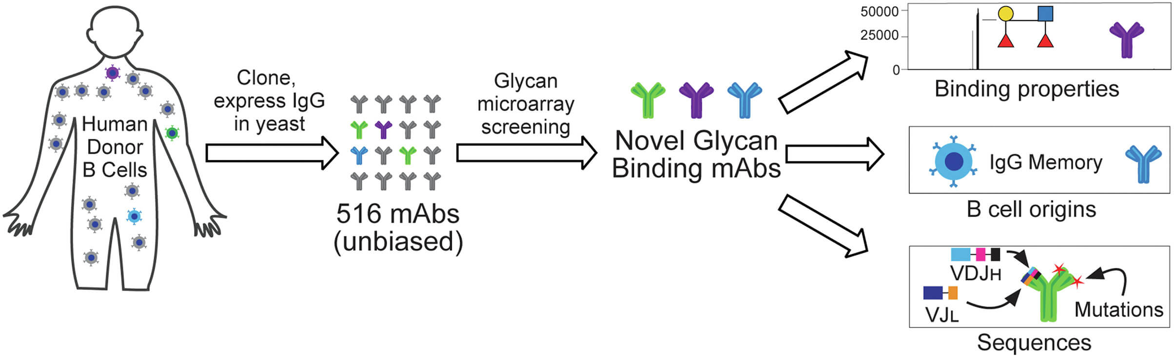
Temme and colleagues used a broad glycan microarray to screen 516 sequence-verified human monoclonal antibodies
derived from multiple B-cell subsets. The array contained more than 800 glycan-related components spanning
diverse carbohydrate classes and presentation contexts. The screen identified 26 antiglycan antibodies, most of
which recognized microbial carbohydrates. Importantly, the majority showed selective binding to defined glycan
motifs rather than broad polyreactivity, demonstrating how large arrays can distinguish focused recognition from
unexpected off-target binding. The study also found that antiglycan antibodies were enriched among IgG⁺ memory
B-cell-derived clones and identified antibodies recognizing glycan structures for which few or no monoclonal
reagents had previously been reported. Together, these results illustrate the value of high-content glycan
arrays for parallel antibody screening, specificity profiling, and candidate prioritization, particularly when
both intended recognition and structurally related cross-reactivity need to be evaluated within the same
experiment.
Fig.3 High-content glycan microarray screening enables parallel identification and specificity
analysis of human glycan-binding monoclonal antibodies.¹
Submit Your Antibody for Glycan Specificity Screening
For a focused project discussion, submit the antibody type and isotype, concentration and buffer,
purification status, target glycan or motif, known positives and negatives, candidate count, desired panel
breadth, project-relevant cross-reactivity concerns, preferred detection route, and the downstream decision
that the screening results will support.
Map My Antibody Glycan
Specificity
Frequently Asked Questions
How does a broad glycan array differ from a targeted panel?
A broad array surveys many glycan classes and is useful when the motif is uncertain or unexpected binding
matters. A compact panel concentrates on structural neighbors around a defined target. The best choice
depends on whether the project is discovering recognition patterns or testing a specific selectivity
boundary.
Can unpurified antibody samples be screened?
Selected hybridoma supernatants, culture supernatants, serum, or plasma may be considered, but matrix
proteins and endogenous antibodies can increase background and complicate attribution. Purified antibody
usually provides clearer interpretation. Sample suitability, dilution, controls, and detection chemistry are
reviewed before the panel and workflow are finalized.
Does a positive array spot prove high affinity?
No. Signal reflects binding under the printed density, orientation, incubation, wash, and detection
conditions. Multivalency can amplify weak interactions, and different probes may present glycans
differently. Affinity or kinetics require an orthogonal method such as SPR or BLI with a suitable analyte
and immobilization design.
How are positive hits selected?
Hit calling considers background, replicate behavior, signal distribution, controls, concentration, and the
purpose of the study. A fixed universal threshold is rarely appropriate across all panels. The analysis plan
sets a suitable level of conservatism and identifies whether borderline signals merit a repeat or focused
follow-up.
Can the array distinguish linkage and positional isomers?
It can compare isomers only when the relevant structures are present as defined probes and the printing
format preserves a meaningful distinction. Absence of differential binding does not prove the antibody
ignores that feature, because density, linker, accessibility, or neighboring structures may mask an
otherwise important difference.
What information is needed to start a screening project?
Provide the antibody form, isotype, species, concentration, purity, buffer, target glycan, expected
near-neighbors, known controls, candidate count, and intended downstream use. Creative Biolabs uses these
details to align panel content, detection, replication, and reporting. For Research Use Only. Not For
Clinical Use.
References
Temme, Joel Sebastian, et al. Microarray-Guided Evaluation of the Frequency, B-Cell Origins, and Selectivity
of Human Glycan-Binding Antibodies Reveals New Insights and Novel Antibodies. Journal of Biological Chemistry 298.10 (2022): 102468. Distributed under Open Access license CC BY 4.0, without modification. https://doi.org/10.1016/j.jbc.2022.102468
Project Inquiry
!For Research Use
Only.Not for clinical or diagnostic use.