- Summarize the project requirements and fill in the information collection form.
- Sign a CDA from both parties to further communicate information, such as targets.
- Select an animal model, discuss experimental design, and determine assay parameters.
- Project costing and project schedule forecasting.
Ischemia-Reperfusion Myocardial Infarction Modeling & Pharmacodynamics Service
At Creative Biolabs, we are acutely aware of these complexities. Leveraging our years of expertise, we offer a comprehensive suite of well-established MI models designed to rigorously evaluate the efficacy of novel cardioprotective agents and therapeutic interventions. Our commitment is to accelerate your research towards impactful solutions.
Introduction
Myocardial Infarction (MI) stands as a formidable global health challenge, exacting a heavy toll in terms of morbidity and mortality. Despite significant advancements in acute treatment, a crucial aspect of post-MI care involves mitigating ischemia-reperfusion (I/R) injury—a paradoxical phenomenon where restoring blood flow to ischemic heart tissue can, ironically, intensify cellular damage. This secondary injury often leads to worsened cardiac function and adverse ventricular remodeling.
Ischemia-Reperfusion Myocardial Infarction Model
At Creative Biolabs, the I/R MI model is established through a sophisticated, highly controlled surgical strategy designed to precisely mimic the transient coronary occlusion and subsequent reperfusion seen in clinical acute MI. Our approach prioritizes meticulous surgical technique combined with advanced micromanipulation to ensure exceptional reproducibility and physiological relevance, which are critical for robust preclinical data generation.
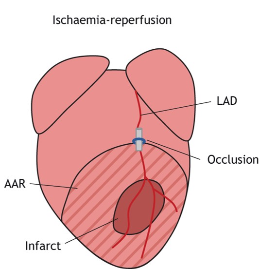 Fig.1 The infarcts generated after ischaemia-reperfusion techniques.1
Model Construction Steps
01Preparation and Anesthesia
Animals (Mouse, Rat, Rabbit, Dog, NHPs) undergo careful preparation, including precise anesthetic induction and maintenance, ensuring stability throughout the procedure. This minimizes stress and variability.
02Thoracotomy and Heart Exposure
A left thoracotomy is performed to carefully expose the heart, providing optimal access to the coronary vasculature.
03LAD Ligation
Under a high-magnification dissecting microscope, the left anterior descending (LAD) coronary artery is identified. A micro-suture is then precisely placed around the LAD and temporarily ligated, inducing controlled myocardial ischemia. Real-time visual changes in the LAD color, coupled with characteristic alterations in electrocardiography (ECG), confirm successful occlusion and ischemia. The duration of ischemia is carefully controlled based on study design.
04Reperfusion
Following the predetermined ischemic period, the micro-suture is carefully released, allowing blood flow to be restored to the ischemic myocardial tissue. This initiation of reperfusion, a critical phase, then allows for the study of I/R injury. ECG monitoring and visual confirmation verify successful reperfusion.
05Post-Surgical Care
Animals receive diligent post-operative care, including pain management and monitoring, to ensure optimal recovery and well-being.
Strengths and Limitations
Strengths:
- Clinical Relevance: The model directly mimics the clinical scenario of MI treated with revascularization, making it highly translatable.
- Study of Reperfusion Injury: It uniquely allows for the investigation of the complex mechanisms and therapeutic targets associated with reperfusion-induced damage, a major contributor to post-MI morbidity.
- Reproducible Infarct Sizes: Highly standardized protocols and skilled technicians ensure consistent infarct sizes, leading to robust and reliable data for compound evaluation.
- Comprehensive Evaluation: The model enables assessment of both acute injury and long-term remodeling, providing a holistic view of therapeutic effects.
Limitations:
- Surgical Complexity: Requires highly skilled surgical expertise and specialized equipment due to the delicate nature of cardiac micro-surgery.
- Invasiveness: As an in vivo surgical model, it is invasive and necessitates stringent ethical considerations and animal welfare protocols.
- Variability (Minimized at Creative Biolabs): While minimized by precision techniques, biological variability inherent in animal models can exist, thereby necessitating robust experimental design and statistical power.
Evaluation Platform
Our state-of-the-art evaluation platform offers a comprehensive array of biochemical, molecular, cellular, histopathological, behavioral, and advanced imaging techniques to rigorously assess therapeutic interventions in the I/R MI model. Our integrated approach ensures deep mechanistic understanding alongside functional outcomes.
Key Test Indicators:
- Biochemical: Cardiac enzymes (e.g., Troponin-I, Troponin-T, CK-MB), inflammatory cytokines (e.g., TNF-α, IL-6), oxidative stress markers (e.g., MDA, SOD).
- Functional Imaging: Echocardiography (e.g., ejection fraction, fractional shortening, left ventricular dimensions), MRI (e.g., infarct size, edema, perfusion), cardiac ultrasound for real-time assessment.
- Histopathology: Infarct size determination (e.g., TTC staining), assessment of cardiomyocyte apoptosis (e.g., TUNEL staining), fibrosis (e.g., Masson's trichrome staining), and inflammatory cell infiltration.
- Molecular/Cellular: Gene expression analysis (qPCR), protein expression (Western Blot, IHC), signaling pathway activation (phosphorylation studies).
- Hemodynamics: Direct measurement of ventricular pressures and cardiac output.
Applications
This versatile model simulates various clinical conditions, primarily acute MI followed by revascularization, and the resultant reperfusion injury. It is extensively utilized to evaluate a wide range of drug classes, including but not limited to:
- Cardioprotective agents: Aiming to preserve myocardial tissue and reduce infarct size.
- Anti-inflammatory compounds: Targeting the detrimental inflammatory response post-I/R.
- Antioxidants: Mitigating oxidative stress-induced damage.
- Agents improving microvascular function: Addressing "no-reflow" phenomena.
- Novel biologics and small molecules: For cardiac regeneration, anti-fibrotic effects, or improvements in cardiac remodeling.
Furthermore, the model facilitates the development and validation of novel therapeutic strategies, such as gene therapies, cell-based therapies, and innovative medical devices, ultimately striving to improve patient outcomes following acute cardiac events.
Related Myocardial Infarction Models
Our Advantages
- Years of Expertise: Deep, specialized knowledge in cardiovascular preclinical models.
- Gold-Standard Models: Meticulously validated and highly reproducible I/R MI models.
- Comprehensive Endpoints: Full suite of functional, histological, biochemical, and molecular analyses.
- Customized Study Design: Flexible protocols tailored precisely to your research objectives.
- Regulatory Alignment: Adherence to ARRIVE guidelines and global ethical standards.
- Accelerated Timelines: Optimized workflows for efficient project completion.
Work with Us
Inquiry Stage
Project Start
- We provide a detailed project plan, including the required sample quantities, methods and protocols.
- Both parties confirm the project details and start the project.
- Confirm the timeline of the project.
Project Progress
- We provide periodic results and information on the animal's condition.
- We will work together to make project adjustments as necessary.
Project Completion
- We provide a comprehensive project report promptly.
- We arrange transportation for the produced samples.
- We provide a discussion of the project results and help to arrange the next steps.
After-Sales Support
- Data storage and archiving.
Contact Us
Partner with Creative Biolabs and leverage our unparalleled expertise in I/R MI models to generate the robust, translational data you need. Contact us today for a personalized consultation or to request a detailed proposal tailored to your specific research objectives.
FAQs
-
Q1: What specific animal species do you use for the I/R MI model?
A: We specialize in ischemia/reperfusion myocardial infarction (I/R MI) models in multiple species, including mice, rats, rabbits, dogs, and non-human primates (NHPs). We can discuss the specific species that best suits your project.
-
Q2: How do you ensure the reproducibility of infarct size in your I/R MI models?
A: Reproducibility is paramount at Creative Biolabs. We achieve consistent infarct sizes through a combination of highly standardized surgical protocols, advanced micromanipulation techniques by highly experienced surgeons, and real-time confirmation using both direct visualization and electrocardiography. Each procedure is meticulously performed to minimize variability.
-
Q3: Can you evaluate both acute and chronic effects of a therapeutic agent in the I/R MI model?
A: Absolutely. Our evaluation platform is equipped to assess both acute cardioprotective effects shortly after reperfusion and long-term impacts on ventricular remodeling, fibrosis, and overall cardiac function over weeks. We design studies with appropriate reperfusion durations and endpoint analyses to capture these different phases of injury and repair.
-
Q4: What quality control measures are in place during the surgical procedure?
A: Quality control is integrated into every step. This includes continuous physiological monitoring (e.g., heart rate, body temperature), real-time visual confirmation of LAD occlusion and reperfusion, and immediate post-surgical animal welfare assessments. Our adherence to ARRIVE guidelines further ensures the ethical and scientific rigor of each experiment.
-
Q5: How do you confirm successful ischemia and reperfusion?
A5: We utilize a dual confirmation approach. Ischemia is visually confirmed by the blanching of the myocardial tissue distal to the LAD ligation and characteristic ST-segment elevation on the electrocardiogram. Reperfusion is similarly confirmed by restoration of blood flow and reversal of ECG changes. This real-time validation is critical for accurate model establishment.
-
Q6: How do you assist with study design and experimental parameters?
A: We offer extensive collaborative support during the study design phase. Our experienced team will work closely with you to define specific research objectives, recommend appropriate experimental parameters (e.g., ischemia/reperfusion durations, animal numbers, endpoints), and help select the most suitable model for your compound's mechanism of action, ensuring a robust and efficient study.
Published Data
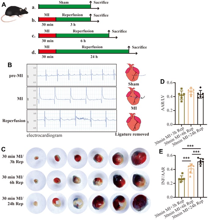 Fig.2 Study on the effect of reperfusion time on infarct size using I/R MI model.2
In a compelling demonstration of the I/R MI model's versatility and its capacity to reveal critical pathophysiological insights, a study investigated the time-dependent phenotypes of myocardial I/R in mice. This research meticulously detailed how varying durations of reperfusion (3, 6, and 24 hours) after 30 or 60 minutes of ischemia led to distinct injury phenotypes. Key findings included an increase in infarct size and apoptosis rates with extended reperfusion, and dynamic changes in serum myocardial enzymes and inflammatory cytokines. This exemplifies how precise control over ischemia and reperfusion parameters allows for the nuanced study of cardiac remodeling and drug effects over different temporal phases.
References
- De Villiers, Carla, and Paul R Riley. "Mouse models of myocardial infarction: comparing permanent ligation and ischaemia-reperfusion." Disease models & mechanisms vol. 13,11 dmm046565. 18 Nov. 2020, DOI:10.1242/dmm.046565. Distributed under Open Access license CC BY 4.0, without modification. The image was modified by extracting and using only part of the original image.
- Meng, Xiang-Min et al. "Evaluation of time-dependent phenotypes of myocardial ischemia-reperfusion in mice." Aging vol. 15,19 (2023): 10627-10639. DOI:10.18632/aging.205103. Distributed under Open Access license CC BY 3.0, without modification.
For Research Use Only.
Online Inquiry