Bilateral Ureteral Obstruction induced Renal Fibrosis Modeling & Pharmacodynamics Service
Creative Biolabs offers a wide range of well-established animal models to evaluate the efficacy of potential therapeutics for CKD. These models include the Bilateral Ureteral Obstruction-Induced Renal Fibrosis Model, 5/6 nephrectomy models, and other CKD-related models. These models are ideal platforms for assessing novel drug candidates targeting renal fibrosis, inflammation, and tissue regeneration, aiding in the development of effective treatments for CKD.
Introduction
Chronic Kidney Disease (CKD) is a progressive condition characterized by a gradual decline in kidney function over time. It is a major global health issue, affecting over 850 million people worldwide. CKD can result from a variety of underlying causes, including diabetes, hypertension, glomerulonephritis, and polycystic kidney disease. As kidney function deteriorates, patients may progress to end-stage renal disease (ESRD), requiring dialysis or kidney transplantation. The progression of CKD is marked by renal fibrosis, which involves the accumulation of extracellular matrix components and the formation of scar tissue, leading to irreversible kidney damage. Early detection and intervention are critical for slowing disease progression and improving patient outcomes. CKD encompasses several stages, with the severity ranging from mild kidney damage (stage 1) to complete kidney failure (stage 5).
Bilateral Ureteral Obstruction-Induced Renal Fibrosis Model
The Bilateral Ureteral Obstruction-Induced Renal Fibrosis Model involves the surgical ligation of both ureters in rodents, resulting in progressive kidney damage and fibrosis due to prolonged renal obstruction. This model effectively mimics the pathological features of CKD, including tubular atrophy, interstitial fibrosis, and glomerulosclerosis. It is commonly used to study the pathophysiology of renal fibrosis, offering a robust platform for testing novel therapeutics targeting fibrosis, inflammation, and kidney injury repair. While highly effective for simulating kidney damage, the model can be associated with a high degree of animal morbidity due to severe obstruction, which may limit long-term survival in some cases. However, the accuracy of renal fibrosis representation makes it indispensable for studying CKD-related fibrosis and evaluating drug efficacy in preclinical stages.
- Simulates: This model primarily simulates chronic kidney disease progression, especially focusing on renal fibrosis induced by bilateral ureteral obstruction.
- Evaluates Drugs: The BUO-induced renal fibrosis model is particularly useful in evaluating therapies aimed at renal fibrosis, fibrosis inhibitors, anti-inflammatory drugs, and agents promoting kidney tissue repair.
Evaluation Platform
- Animals: Mouse, Rat.
-
Measurements
We offer a comprehensive suite of measurement techniques to evaluate drug efficacy in the Bilateral Ureteral Obstruction-Induced Renal Fibrosis Model, including but not limited to:- General observations: body weight, mortality rate, renal function (creatinine, blood urea nitrogen levels).
- Histological analysis: tissue sections stained for collagen deposition, tubular damage, and inflammatory cell infiltration.
- Immunohistochemistry: evaluation of fibrosis markers (e.g., α-SMA, collagen I, TGF-β) and immune cell infiltration (e.g., macrophages, T-cells).
- Cytokine profiling (e.g., ELISA): levels of pro-inflammatory cytokines such as IL-6, TNF-α, and IL-1β.
- Gene/protein expression profiling: RT-qPCR and Western blot analysis for fibrosis-related genes (e.g., Col1a1, TGF-β1, FN1).
Our scientific team is ready to assist with experimental design, model selection, and data interpretation, ensuring that each project is customized to meet your specific research needs.
Related Services
In addition to the Bilateral Ureteral Obstruction-Induced Renal Fibrosis Model, we offer other animal models for chronic kidney disease. These models can be used to evaluate a wide range of therapeutic strategies targeting renal injury and fibrosis.
- Unilateral Ureter Obstruction (UUO) Model
- 5/6 Nephrectomy Model
- Adriamycin induced Nephropathy (AN) Rodent Model
- Folic Acid (FA) induced Renal Fibrosis Model
- Adenine induced Chronic Renal Failure Model
Our advantages
- Comprehensive Services: From model selection and design to data analysis, we provide end-to-end services.
- Expert Scientific Team: Our experienced team ensures accurate results, guiding you through each phase of your research.
- Tailored Solutions: We adapt our services to your specific research needs, delivering customized protocols and analyses.
- High Reproducibility: The models we provide have been validated for reproducibility and high-quality data generation.
- Cutting-Edge Technologies: Access to the latest advancements in molecular and histological techniques to enhance the precision of your results.
- Regulatory Compliance: We ensure all our models and data adhere to the highest ethical and regulatory standards for research.
Work with Us
- Summarize the project requirements and fill in the information collection form.
- Sign a CDA from both parties to further communicate information, such as targets.
- Select an animal model, discuss experimental design, and determine assay parameters.
- Project costing and project schedule forecasting.
- We provide a detailed project plan, including the required sample quantities, methods, and protocols.
- Both parties confirm the project details and start the project.
- Confirm the timeline of the project.
- We provide periodic results and information on the animal's condition.
- We will work together to make project adjustments as necessary.
- We provide a comprehensive project report promptly.
- We arrange transportation for the produced samples.
- We provide a discussion of the project results and help to arrange the next steps.
- Data storage and archiving.
FAQs
-
1. What is the advantage of using the Bilateral Ureteral Obstruction-Induced Renal Fibrosis Model in CKD research?
This model closely mimics the pathological progression of renal fibrosis and obstructive nephropathy, providing a reliable platform for testing therapeutic interventions.
-
2. How long does it take to develop renal fibrosis in this model?
Renal fibrosis typically begins to develop after 2-3 weeks post-surgery, with more pronounced fibrosis observed after 4-6 weeks.
-
3. Are there alternative methods to assess renal fibrosis?
Yes, we also offer other models, such as the 5/6 nephrectomy model and Adriamycin-induced nephropathy model, which may be suitable depending on the specific research goals.
-
4. What types of drugs can be tested using this model?
The model is ideal for testing anti-fibrotic drugs, kidney regeneration therapies, anti-inflammatory agents, and drugs targeting renal fibrosis pathways.
-
5. Can I customize the protocol for my specific research needs?
Absolutely! We provide flexible protocols tailored to meet your unique experimental requirements, ensuring the most relevant data for your study.
Published Data
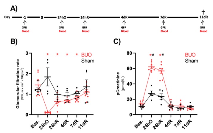 Fig. 1 The impact of bilateral ureteral obstruction on tGFR and pCreatinine.1
The study aimed to assess the applicability of transcutaneous glomerular filtration rate (tGFR) measurements for monitoring renal function changes. The impact of bilateral ureteral obstruction (BUO) and subsequent release on GFR was evaluated, as illustrated in Figure 1A. As anticipated, Figure 1B shows a significant decrease in GFR 24 hours post-BUO, while Sham animals did not exhibit similar changes. Following the release of the obstruction, the GFR in the BUO group remained consistently lower than baseline levels, although after 11 days, it approached baseline values and was no longer significantly different. Plasma creatinine (pCreatinine) levels were markedly elevated 24 hours after BUO and 24 hours post-release; however, by day 4, pCreatinine levels returned to baseline levels (Figure 1C). A similar, though less pronounced, effect was observed in the Sham group (Figure 1C).
Reference
- Jensen, Michael Schou et al. "Transcutaneous measurement of renal function in two rodent models of obstructive nephropathy." BMC research notes vol. 16,1 119. 26 Jun. 2023. Distributed under Open Access license CC BY 4.0, without modification. https://doi.org/10.1186/s13104-023-06387-y
For Research Use Only.