Streptozotocin (STZ) & High-Fat induced Non-Alcoholic Steatohepatitis (NASH) Modeling & Pharmacodynamics Service
Creative Biolabs offers a wide range of preclinical models to evaluate the efficacy of NASH therapeutics, providing reliable, accurate, and reproducible services that support drug development at various stages.
Introduction
Non-alcoholic steatohepatitis (NASH) is a chronic liver disease characterized by inflammation, hepatocyte injury, and progressive fibrosis, which can lead to cirrhosis, liver failure, or hepatocellular carcinoma. It is part of the spectrum of non-alcoholic fatty liver disease (NAFLD), which encompasses a range of liver abnormalities caused by metabolic risk factors, such as obesity, diabetes, and dyslipidemia. NASH is becoming increasingly prevalent globally, driven by the rising rates of metabolic disorders. The pathogenesis of NASH involves complex mechanisms, including insulin resistance, oxidative stress, inflammatory cytokines, and altered lipid metabolism. Early diagnosis is crucial for effective intervention and prevention of disease progression. Imaging, liver biopsy, and biochemical markers are commonly used in clinical practice to assess the severity of liver damage.
Disease Models and Applications
The Streptozotocin (STZ) & High-Fat induced NASH Model is commonly used to simulate human NASH. This model is constructed by administering STZ to induce insulin resistance and a high-fat diet to induce steatosis. STZ works by selectively damaging pancreatic beta cells, leading to hyperglycemia, while the high-fat diet mimics the lipid accumulation in hepatocytes characteristic of NASH. The model displays a range of key NASH features, such as hepatic inflammation, steatosis, and fibrosis. The advantages of this model include its ability to replicate the pathophysiology of human NASH, making it suitable for drug testing. However, it has limitations, such as the artificial nature of the STZ induced diabetes and the variability in response depending on the species used.
- Simulates: The Streptozotocin (STZ) & High-Fat induced NASH Model simulates NASH, specifically focusing on liver steatosis, inflammation, and fibrosis. It helps researchers understand the progression of non-alcoholic fatty liver disease (NAFLD) to NASH and study the effects of therapeutic interventions.
- Evaluates Drugs: This model is used to evaluate drugs that target multiple stages of NASH, including those aimed at reducing liver fat accumulation (e.g., PPAR agonists), anti-inflammatory agents (e.g., TNF-α inhibitors), and antifibrotic drugs. It is also valuable for testing insulin-sensitizing agents or drugs that aim to restore normal liver function and structure.
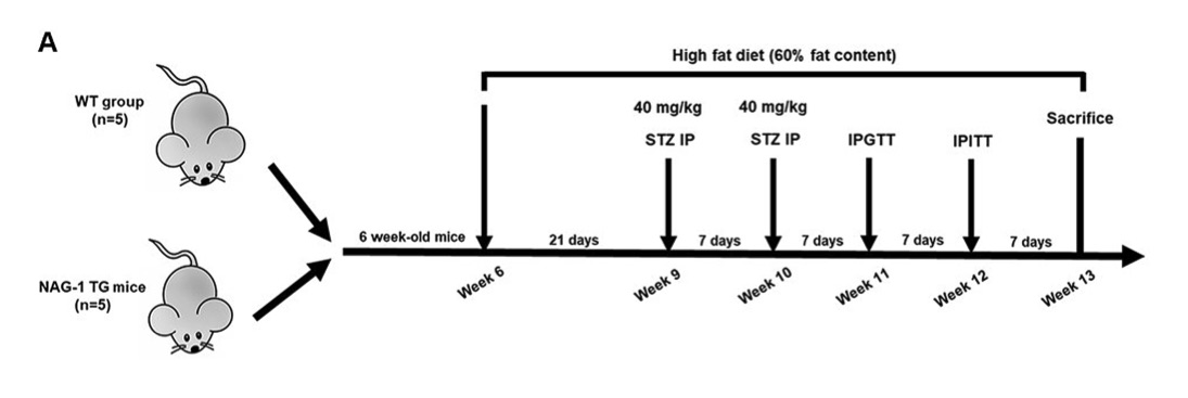 Fig. 1 Schematic diagram representing the timeline for experiments using the diabetes mouse model.1
Measurements
We offer a variety of measurements for evaluating drug efficacy in rodent NASH models, utilizing an array of advanced technologies, including but not limited to:
- General Observations: Body weight, mortality rate, stool consistency, gastrointestinal bleeding.
- Histopathology: Liver tissue analysis via H&E staining to assess steatosis, inflammation, and fibrosis.
- Immunohistochemistry: Infiltration of immune cells (e.g., T-cells, macrophages) in liver tissues.
- Cytokine Profiling (e.g., ELISA): Expression levels of inflammatory mediators such as TNF-α, IL-6, and IL-1β.
- Hematology Analysis and Serum Biomarkers: Liver enzymes, bilirubin levels, and other serum markers of liver injury.
- Gene/Protein Expression Profiling: RT-qPCR and Western blot techniques to assess expression levels of liver fibrosis markers (e.g., collagen, α-SMA).
In addition to the established NASH models, our expertise extends to the development of novel animal models tailored to specific research needs. Our scientific team is available to assist in experimental design, model selection, and data analysis, ensuring a customized and effective approach to your project.
Related Services
Aside from the Streptozotocin (STZ) & High-Fat induced NASH Model, we also offer other NASH models. These alternative approaches provide additional insights into the disease and allow for testing a broader range of therapeutic strategies.
- Diet induced Obesity (DIO) Mouse NASH Model
- High-Fat Diet induced NASH Model
- Methionine Choline-Deficient (MCD) Diet induced NASH Model
- Choline-Deficient L-Amino Acid-Defined (CDAA) Diet induced NASH Model
- High-Fat & High-Carbohydrate Diet induced NASH Model
- High-Fat & High-Cholesterol Diet induced NASH Model
- High-Fat & High-Cholesterol Diet & Fructose induced NASH Model
- High-Fat & Fructose induced NASH Model
- Diethylnitrosamine (DEN) & High-Fat & High-Carbohydrate Diet induced NASH Model
- High-Fat & CCL4 induced NASH Model
- MC4R KO Mouse Model
- LDLR KO Mouse Model
Advantages
- Experienced Scientific Team: Our experts provide consultation, model development, and data analysis tailored to your research needs.
- Customized Solutions: We design and develop models suited to specific therapeutic targets, enhancing the relevance of your research.
- Comprehensive Services: From model creation to drug evaluation and biomarker analysis, we provide end-to-end support.
- Advanced Technology: We utilize state-of-the-art technologies for precise and accurate measurements of therapeutic efficacy.
- Reliability and Precision: Our models have been validated in numerous studies, ensuring high reproducibility and reliability of results.
Work with Us
- Summarize the project requirements and fill in the information collection form.
- Sign a CDA from both parties to further communicate information, such as targets.
- Select an animal model, discuss experimental design, and determine assay parameters.
- Project costing and project schedule forecasting.
- We provide a detailed project plan, including the required sample quantities, methods, and protocols.
- Both parties confirm the project details and start the project.
- Confirm the timeline of the project.
- We provide periodic results and information on the animal's condition.
- We will work together to make project adjustments as necessary.
- We provide a comprehensive project report promptly.
- We arrange transportation for the produced samples.
- We provide a discussion of the project results and help to arrange the next steps.
- Data storage and archiving.
FAQs
-
Q: What is NASH, and how is it different from NAFLD?
A: NASH is a more advanced form of NAFLD, characterized by liver inflammation and fibrosis. While NAFLD involves fat accumulation without inflammation, NASH progresses with liver injury and scarring, potentially leading to cirrhosis.
-
Q: How long does it take to establish a Streptozotocin (STZ) & High-Fat induced NASH model?
A: It typically takes around 12-16 weeks to induce and monitor NASH in this model, including time for drug treatment and evaluation.
-
Q: What types of drugs can be tested using this model?
A: This model can be used to evaluate drugs targeting fat accumulation, inflammation, and fibrosis, including PPAR agonists, anti-inflammatory agents, and antifibrotic therapies.
-
Q: What are the key biomarkers measured in this model?
A: Key biomarkers include liver enzymes, cytokines (e.g., TNF-α, IL-6), fibrosis markers (e.g., collagen), and gene/protein expression related to liver injury.
-
Q: Can this model be used for both prevention and treatment studies?
A: Yes, this model is versatile and can be used to evaluate both preventive interventions and treatments for established NASH.
Published Data
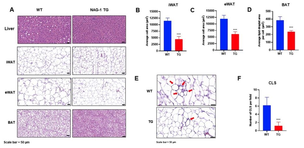 Fig. 2 The effect of curcumin intervention on histopathological changes in the livers of rats fed a high-fat diet then treated with streptozotocin.1
NAG-1 overexpression was shown to attenuate high-fat diet/streptozotocin induced diabetic dyslipidemia and hepatic steatosis in mice. Tissue sections from the liver, inguinal white adipose tissue (iWAT), epididymal white adipose tissue (eWAT), and brown adipose tissue (BAT) were prepared and stained with hematoxylin and eosin (H&E). Histological images of these tissues are presented in Fig. 2A. In the liver, lipid accumulation and the number of fat droplets were reduced in NAG-1 Tg mice compared to wild-type (WT) mice, indicating a decrease in hepatic steatosis upon NAG-1 overexpression. Additionally, hypertrophy of adipose tissue was less pronounced in NAG-1 Tg mice than in WT mice. Quantification of the histological data revealed that the size of lipid droplets in iWAT, eWAT, and BAT was significantly smaller in NAG-1 Tg mice compared to WT mice (p < 0.001) (Fig. 2B–D). Furthermore, the number of crown-like structures (CLS) in eWAT, a histological marker of pro-inflammatory processes in adipose tissue, was significantly lower in NAG-1 Tg mice than in WT mice (p < 0.001), suggesting a reduction in inflammatory lesions (Fig. 2E, F).
Reference
- Lertpatipanpong, Pattawika et al. "The anti-diabetic effects of NAG-1/GDF15 on HFD/STZ induced mice." Scientific Reports vol. 11,1 15027. 22 Jul. 2021, DOI:10.1038/s41598-021-94581-y. Distributed under an Open Access license CC BY 4.0, without modification.
For Research Use Only.