Methionine Choline-Deficient (MCD) Diet induced Non-Alcoholic Steatohepatitis (NASH) Modeling & Pharmacodynamics Service
Creative Biolabs provides a variety of well-established models to evaluate the efficacy of drugs targeting NASH, offering reliable platforms for preclinical testing and therapeutic development for liver diseases.
Introduction
Non-alcoholic steatohepatitis (NASH) is a progressive liver disease characterized by the accumulation of fat in liver cells, inflammation, hepatocellular injury, and varying degrees of fibrosis. It is considered a more severe form of non-alcoholic fatty liver disease (NAFLD) and is often associated with metabolic conditions such as obesity, type 2 diabetes, and hyperlipidemia. NASH is recognized as a leading cause of cirrhosis, liver failure, and hepatocellular carcinoma. The pathogenesis of NASH involves a complex interplay of lipid accumulation, oxidative stress, inflammatory cytokine release, and hepatic fibrosis. As the global prevalence of obesity and metabolic syndrome continues to rise, NASH has become an urgent public health issue. The disease progresses through several stages, beginning with simple hepatic steatosis and, if left untreated, advancing to fibrosis and eventually cirrhosis or liver cancer. Early detection and therapeutic intervention are critical in preventing the progression of NASH and improving patient outcomes. Currently, there are no FDA-approved therapies specifically for NASH, making it a significant focus of research and drug development.
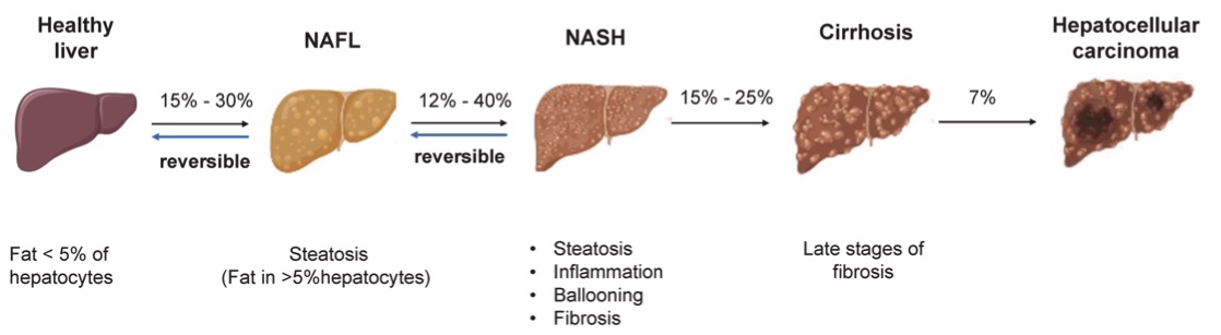 Fig. 1 The pathogenesis of NAFLD.1
Disease Models and Applications
The Methionine Choline-Deficient (MCD) Diet induced NASH Model is widely used to replicate the pathological features of NASH in rodents. This model is established by feeding rodents an MCD diet, which lacks methionine and choline, essential nutrients for proper liver function. The MCD diet induces a rapid accumulation of fat in hepatocytes, leading to liver injury, inflammation, and fibrosis, closely mimicking the progression of human NASH. The key advantage of this model is its ability to produce a severe form of NASH with significant fibrosis and hepatocellular injury within a relatively short period. However, it has some limitations, such as the absence of obesity, which is commonly observed in human NASH, and the fact that the MCD diet can also lead to hepatic damage through mechanisms not typically associated with metabolic syndrome. Despite these limitations, the MCD induced NASH model is a reliable tool for evaluating drug candidates targeting liver inflammation, fibrosis, and metabolic dysfunction.
- Simulates: The Methionine Choline-Deficient (MCD) Diet induced NASH Model simulates non-alcoholic steatohepatitis with significant liver injury, inflammation, and fibrosis in rodents. It closely mimics the hepatic inflammation and fibrosis observed in human NASH, making it a valuable model for studying disease progression and testing therapeutic interventions.
- Evaluates Drugs: This model is commonly used to evaluate drugs aimed at reducing liver fat accumulation, inflammation, oxidative stress, and fibrosis. Drug candidates tested in this model include anti-inflammatory agents, antioxidants, antifibrotic compounds, and metabolic regulators that may prevent the progression of NASH to cirrhosis or liver cancer.
Measurements
We offer a variety of measurements for evaluating drug efficacy in the Methionine Choline-Deficient (MCD) Diet induced NASH Model, utilizing advanced technologies, including but not limited to:
- General observations: body weight, liver weight, and liver enzyme levels (e.g., ALT, AST, GGT) in serum to assess liver function.
- Histopathology: Liver tissue examination for steatosis, inflammation, and fibrosis using standard stains such as H&E (Hematoxylin and Eosin) and Masson's trichrome (to detect collagen deposition).
- Immunohistochemistry: Analysis for inflammation and fibrosis markers, including TNF-α, IL-6, α-SMA, and collagen I, to evaluate the degree of liver injury and fibrosis.
- Cytokine profiling (e.g., ELISA): Quantification of pro-inflammatory cytokines such as TNF-α, IL-1β, IL-6, and MCP-1 to assess inflammation levels in liver tissues.
- Gene/protein expression profiling: RT-qPCR and Western blot techniques to measure genes and proteins involved in inflammation (e.g., NF-kB, iNOS), fibrosis (e.g., TGF-β, α-SMA), and lipid metabolism (e.g., PPAR-γ, SREBP-1c).
Additionally, our scientific team provides guidance on experimental design, model selection, and data analysis to ensure the success of your research project.
Related Services
In addition to the Methionine Choline-Deficient (MCD) Diet induced NASH Model, we offer other models for inducing NASH. These alternative methods can be used to assess different aspects of NASH pathology and therapeutic interventions.
- Diet induced Obesity (DIO) Mouse NASH Model
- High-Fat Diet induced NASH Model
- Choline-Deficient L-Amino Acid-Defined (CDAA) Diet induced NASH Model
- High-Fat & High-Carbohydrate Diet induced NASH Model
- High-Fat & High-Cholesterol Diet induced NASH Model
- High-Fat & High-Cholesterol Diet & Fructose induced NASH Model
- High-Fat & Fructose induced NASH Model
- Diethylnitrosamine (DEN) & High-Fat & High-Carbohydrate Diet induced NASH Model
- High-Fat & CCL4 induced NASH Model
- Streptozotocin (STZ) & High-Fat induced NASH Model
- MC4R KO Mouse Model
- LDLR KO Mouse Model
Advantages
- Comprehensive Expertise: Our team has extensive experience in liver disease modeling, offering tailored solutions for your specific research needs.
- Customizable Models: We offer flexible options for customizing NASH models, including adjusting diet compositions, drug dosing, and experimental protocols to meet your study requirements.
- Validated and Reliable Models: Our models are validated based on the latest scientific literature and are proven to closely mimic human disease progression, ensuring the reliability of your preclinical data.
- Advanced Technologies: We use state-of-the-art tools such as histology, immunohistochemistry, and gene expression profiling to provide detailed and accurate assessments of drug efficacy.
- End-to-End Support: From model selection to data analysis, our team offers complete support throughout your research, ensuring the best possible outcomes for your project.
- Global Reach: We serve research institutions, pharmaceutical companies, and biotech firms worldwide, providing comprehensive services for drug discovery and development.
Work with Us
- Summarize the project requirements and fill in the information collection form.
- Sign a CDA from both parties to further communicate information, such as targets.
- Select an animal model, discuss experimental design, and determine assay parameters.
- Project costing and project schedule forecasting.
- We provide a detailed project plan, including the required sample quantities, methods, and protocols.
- Both parties confirm the project details and start the project.
- Confirm the timeline of the project.
- We provide periodic results and information on the animal's condition.
- We will work together to make project adjustments as necessary.
- We provide a comprehensive project report promptly.
- We arrange transportation for the produced samples.
- We provide a discussion of the project results and help to arrange the next steps.
- Data storage and archiving.
FAQs
-
Q: How long does it take to induce NASH in the MCD diet model?
A: It typically takes 4-6 weeks of MCD diet feeding to observe significant hepatic steatosis, inflammation, and fibrosis in rodents.
-
Q: Can the MCD diet model be used to evaluate antifibrotic therapies?
A: Yes, the MCD diet induced NASH model is widely used to evaluate antifibrotic therapies, as it induces liver fibrosis similar to human NASH.
-
Q: Are there any limitations to using the MCD diet model?
A: The MCD diet model lacks the obesity component typically seen in human NASH, and it may not fully replicate all aspects of human metabolic syndrome. However, it remains an essential tool for studying liver injury and fibrosis.
-
Q: Do you offer assistance with experimental design?
A: Yes, our scientific team is available to assist with experimental design, model selection, and data interpretation to ensure the success of your research project.
Published Data
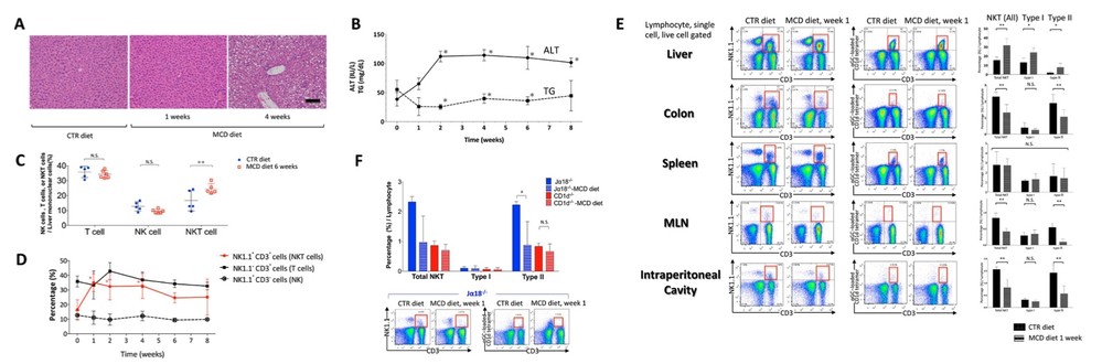 Fig. 2 Methionine-choline deficient (MCD) diet induces nonalcoholic steatohepatitis (NASH) and natural killer T (NKT) cell accumulation.2
Mice fed the MCD (Methionine Choline-Deficient) diet for 4 weeks displayed significant steatohepatitis and increased macroscopic lipid content compared to control (CTR)-fed mice (Fig. 2A). From week 2 to week 8, MCD mice showed elevated levels of alanine aminotransferase (ALT) and decreased triglyceride levels (Fig. 2B). The immune cell subsets in the liver were also analyzed in these mice. Consistent with previous studies, the number of hepatic NK1.1+CD3+ T cells (both type I and type II NKT cells) progressively increased from weeks 1 to 6 under the MCD diet (Fig. 2C and 2D). Interestingly, the ratio of NK1.1- CD3+ T cells (T cells) to NK1.1+ CD3- cells (NK cells) remained unchanged through week 8 of the MCD diet (Fig. 2D). These findings suggest that choline deficiency specifically impacts hepatic NKT cells. Additionally, the proportion of hepatic macrophages, dendritic cells (DCs), and Gr-1+ cells increased, aligning with previous reports. A notable observation was that significantly fewer NKT cells were found in the colonic lamina propria, spleen, mesenteric lymph nodes, and peritoneal cavity compared to the liver of MCD mice, which also mirrors prior findings. In the liver of MCD mice, both type I and type II NKT cell numbers increased, while type II NKT cells were significantly reduced in the colonic lamina propria, mesenteric lymph nodes, and peritoneal cavity compared to CTR mice. Furthermore, while the number of NKT cells without the α-GalCer-loaded Cd1d tetramer (type II NKT cells) was reduced in the colonic lamina propria after 1 week of MCD feeding, type I NKT cells remained unaffected (Fig. 2E). Analysis of NKT cell frequencies in colonic lamina propria of type I NKT cell-deficient (Jα18-/-) and type I and type II NKT cell-deficient (CD1d-/-) mice revealed a significant reduction only in type II NKT cells (Fig. 2F). These results indicate that choline deficiency leads to a decrease in type II NKT cell accumulation in the colonic lamina propria.
References
- Fang, Tingyu et al. "Mouse models of nonalcoholic fatty liver disease (NAFLD): pathomechanisms and pharmacotherapies." International Journal of Biological Sciences vol. 18,15 5681-5697. 6 Sep. 2022, DOI:10.7150/ijbs.65044. Distributed under an Open Access license CC BY 4.0, without modification.
- Sagami, Shintaro et al. "Choline Deficiency Causes Colonic Type II Natural Killer T (NKT) Cell Loss and Alleviates Murine Colitis under Type I NKT Cell Deficiency." PloS one vol. 12,1 e0169681. 17 Jan. 2017, DOI: 10.1371/journal.pone.016968. Distributed under an Open Access license CC BY 4.0, without modification.
For Research Use Only.