Carotid Artery Endothelial Denudation Modeling & Pharmacodynamics Service
At Creative Biolabs, we are dedicated to advancing this understanding by providing a variety of well-established and highly relevant in vivo models for evaluating the efficacy of novel anti-atherosclerotic interventions.
Introduction
Atherosclerosis, a chronic inflammatory disease of the arteries, is a primary driver of cardiovascular morbidity and mortality worldwide. Characterized by the buildup of plaques within arterial walls, it leads to arterial narrowing, reduced blood flow, and severe complications like heart attack and stroke. Understanding the intricate mechanisms of atherosclerosis and developing effective therapeutic strategies is paramount for global health.
Carotid Artery Endothelial Denudation Model
The carotid artery endothelial denudation model is an indispensable in vivo tool for studying vascular injury, restenosis, and early atherosclerosis. By precisely removing the carotid artery's endothelial lining, it acutely initiates biological responses mimicking human pathologies like those post-angioplasty. This robust platform investigates intimal hyperplasia, smooth muscle cell proliferation, matrix remodeling, and neointimal lesion formation. It is invaluable for evaluating therapeutics preventing vascular remodeling, assessing endothelial functional recovery, and exploring the interplay between endothelial integrity, shear stress, and atherosclerotic plaque formation.
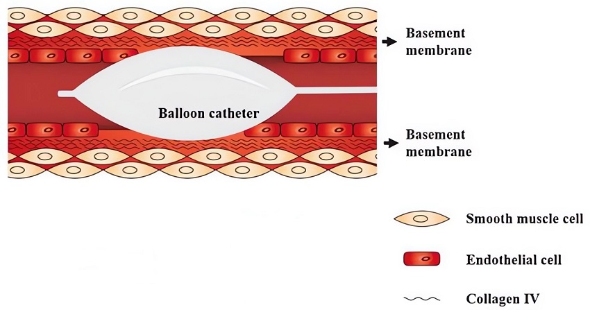 Fig.1 The balloon angioplasty for endothelial denudation of the carotid artery.1
Model Construction Steps
The model is built via a precise surgical procedure. Its core strategy involves mechanically removing the endothelium from a defined segment of the common carotid artery in rodents using a balloon catheter. This ensures reproducible injury while preserving the underlying smooth muscle layer.
01Anesthesia and Surgical Exposure
The experimental animal (e.g., rat or mouse) is anesthetized, and a sterile surgical field is prepared. The common carotid artery is carefully exposed through a small incision.
02Catheter Insertion
A small arteriotomy is performed, usually in the external carotid artery. A balloon catheter, specifically sized for the vessel, is then gently inserted into the common carotid artery.
03Endothelial Denudation
The balloon is advanced to the desired segment, inflated with a precise volume of air or saline for a controlled duration, and then withdrawn slowly several times along the vessel lumen. This action mechanically denudes the endothelium.
04Ligation and Closure
After denudation, the catheter is removed, and the arteriotomy site is ligated. The surgical incision is then closed, and the animal is allowed to recover.
05Post-Operative Care
Animals receive appropriate analgesia and monitoring to ensure proper recovery and minimize stress.
Strengths and Limitations
Strengths:
- Physiological Relevance: Accurately mimics mechanical vascular injury seen in human conditions like angioplasty and stent placement.
- High Reproducibility: Standardized protocols ensure consistent injury and reliable, comparable results across experiments.
- Quantifiable Endpoints: Allows for precise measurement of neointimal formation, lumen diameter, cell proliferation, and functional vascular responses.
- Versatility: Suitable for time-course studies, drug efficacy testing, medical device evaluation, and investigating disease mechanisms.
- Genetic Compatibility: Easily combined with genetically modified animal models to explore gene-specific effects.
- Proof-of-Concept: An ideal model for initial proof-of-concept investigations for both therapeutic and diagnostic innovations.
Limitations:
- Acute Injury Model: Primarily represents an acute injury response rather than chronic, spontaneous atherosclerosis development.
- Single Vessel Focus: Typically involves injury to a single carotid artery, which may not fully reflect systemic vascular disease.
- Surgical Expertise: Requires highly skilled surgical teams to ensure consistent and successful denudation.
Evaluation Platform
Creative Biolabs' comprehensive evaluation platform supports in-depth analysis of the model, utilizing state-of-the-art instruments for:
- Morphometric Analysis: Precise measurements of intimal area, medial area, lumen area, and intimal-to-medial ratio.
- Cellular Analysis: Quantification of cell proliferation markers (e.g., PCNA, Ki67) and inflammatory cell infiltration.
- Molecular Analysis: Assessment of extracellular matrix composition.
- Functional Assessments: Evaluation of vascular reactivity and re-endothelialization.
- Imaging: Advanced imaging techniques for detailed visual and quantitative data.
Applications
- Disease Modeling and Mechanistic Studies: This model accurately simulates key vascular pathologies like restenosis, intimal hyperplasia, and early atherosclerosis, providing a dynamic in vivo platform to unravel the underlying cellular and molecular mechanisms of vascular injury and remodeling.
- Therapeutic and Device Evaluation: It is extensively used to evaluate novel pharmacological agents (e.g., anti-restenotic, anti-inflammatory, pro-endothelial repair drugs) and assess the efficacy and biocompatibility of medical devices, including stent coatings and drug-eluting balloons.
- Diagnostic and Biomarker Development: The model is instrumental in identifying and validating new biomarkers for vascular injury and inflammation, as well as in developing and testing advanced diagnostic techniques for early detection of vascular pathologies.
Related Atherosclerosis Models
- ApoE-/- Mice Model
- Low-Density Lipoprotein Receptor-Deficient Mice (LDLR-/-) Model
- ApoE*3 Transgenic (E3L) Mice Model
- Fatty Zucker Rats Model
- High-Fat-Diet (HFD) & CHOL-Induced Aorta Atherosclerosis Model
- Blood Flow-Induced Arterial Intimal Thickening Model
Our Advantages
- Extensive Experience: Years of expertise in preclinical models, ensuring reliable and high-quality data.
- State-of-the-Art Facilities: Access to advanced surgical suites, imaging technologies, and analytical capabilities.
- Expert Team: Highly skilled scientific and surgical teams dedicated to precise model execution and data interpretation.
- Customized Study Design: Tailored protocols developed in close collaboration to meet your specific research objectives.
- Comprehensive Data Delivery: Provision of high-quality, interpretable data packages, accelerating your research and development.
Work with Us
- Summarize the project requirements and fill in the information collection form.
- Sign a CDA from both parties to further communicate information, such as targets.
- Select an animal model, discuss experimental design, and determine assay parameters.
- Project costing and project schedule forecasting.
- We provide a detailed project plan, including the required sample quantities, methods, and protocols.
- Both parties confirm the project details and start the project.
- Confirm the timeline of the project.
- We provide periodic results and information on the animal's condition.
- We will work together to make project adjustments as necessary.
- We provide a comprehensive project report promptly.
- We arrange transportation for the produced samples.
- We provide a discussion of the project results and help to arrange the next steps.
- Data storage and archiving.
Contact Us
Creative Biolabs provides comprehensive preclinical services utilizing the carotid artery endothelial denudation model, designed to accelerate your cardiovascular drug and device development. We invite you to contact us to discuss how our specialized expertise can support your research goals and contribute to your next scientific success.
FAQs
-
Q1: How does this model mimic human vascular disease?
A: The model closely replicates the acute vascular injury seen in patients undergoing interventional procedures like angioplasty. The subsequent biological responses, including inflammation, smooth muscle cell proliferation, and neointimal formation, are highly similar to those observed in human restenosis and contribute to the progression of atherosclerosis.
-
Q2: What are the key endpoints measured in studies using this model?
A: Researchers typically measure several key endpoints to assess the extent of vascular remodeling and the efficacy of interventions. These include morphometric analyses such as neointimal area, lumen diameter, and the ratio of intimal to medial thickness. Additionally, cellular proliferation markers and inflammatory cell infiltration are often quantified.
-
Q3: Can this model be used to evaluate both drugs and medical devices?
A: Absolutely. The model is highly versatile and serves as an excellent platform for evaluating both novel pharmacological agents and medical devices. For drugs, it helps assess their anti-restenotic or anti-inflammatory properties. For devices, it allows for the evaluation of biocompatibility and their impact on vascular healing and long-term patency.
-
Q4: What are the advantages of using this model over in vitro studies?
A: While in vitro studies provide valuable mechanistic insights, the in vivo model offers a more physiologically relevant environment. It accounts for complex interactions between various cell types, systemic factors, and hemodynamic forces that cannot be fully replicated in a dish, providing more translatable results.
-
Q5: How reproducible are the results obtained from this model?
A: When performed by experienced surgical teams following standardized protocols, the model yields highly reproducible results. This consistency is crucial for generating reliable data, minimizing experimental variability, and ensuring the statistical power necessary for robust scientific conclusions.
-
Q6: Does Creative Biolabs offer customized study designs for this model?
A: Yes, Creative Biolabs specializes in providing tailored research solutions. We collaborate closely with our clients to design studies that precisely meet their specific research objectives. This collaborative approach ensures that the experimental design is optimized to address unique scientific questions and deliver meaningful outcomes.
Published Data
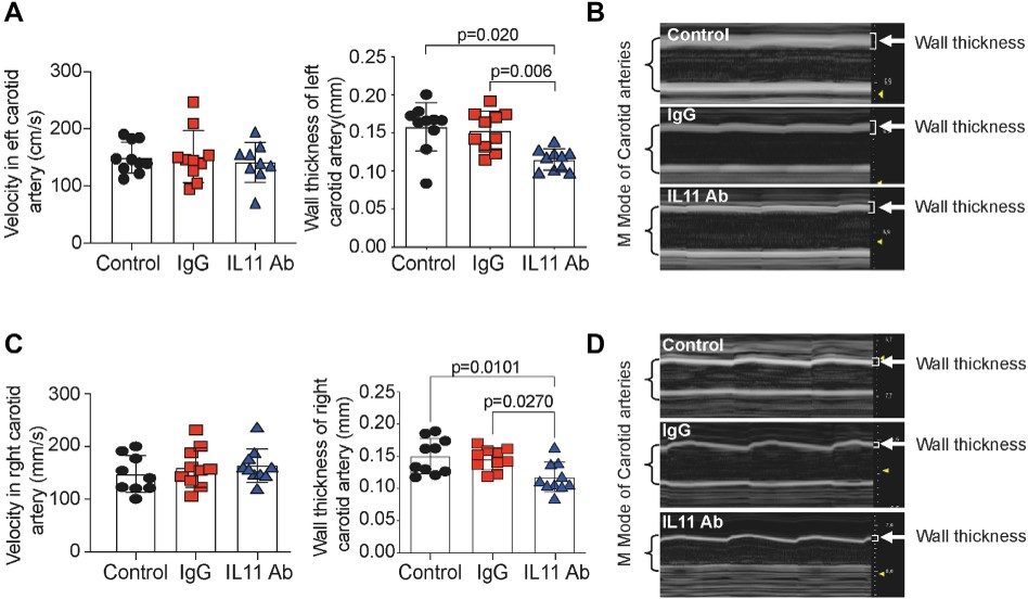 Fig.2 Impact of anti-IL-11 antibody treatment on vessel wall thickness.2
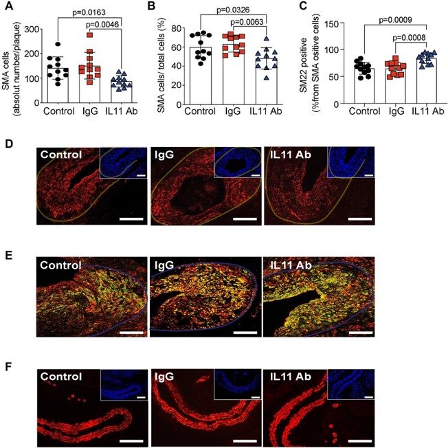 Fig.3 Impact of anti-IL-11 antibody treatment on injury-related plaque VSMC accumulation.2
This research demonstrated that inhibiting Interleukin 11 (IL-11) with a neutralizing antibody significantly reduced post-endothelial injury vessel wall thickness and attenuated neointimal hyperplasia. Mitigating these pathological changes, IL-11 inhibition emerges as a novel therapeutic approach to reduce arterial stenosis post-revascularization, offering a new avenue for preventing artery re-narrowing. The carotid artery endothelial denudation model's effectiveness in identifying and validating novel therapeutic targets and its precise in vivo quantification capabilities make it indispensable for advancing cardiovascular drug discovery.
References
- Mo, Xinhai et al. "Molecular Ultrasound Monitoring of Early Artery Injury After Carotid Balloon Angioplasty." Frontiers in pharmacology vol. 9 1569. 25 Jan. 2019. Distributed under Open Access license CC BY 4.0, without modification. The image was modified by extracting and using only part of the original image. https://doi.org/10.3389/fphar.2018.01569
- Schumacher, David et al. "A neutralizing IL-11 antibody reduces vessel hyperplasia in a mouse carotid artery wire injury model." Scientific reports vol. 11,1 20674. 19 Oct. 2021. Distributed under Open Access license CC BY 4.0, without modification. https://doi.org/10.1038/s41598-021-99880-y
For Research Use Only.